- *Corresponding Author:
- Haiying Dong
Department of Pathology
School of Pathology, Qiqihar Medical University
Qiqihar, Heilongjiang 161006, China
E-mail: dd6457@126.com
| This article was originally published in a special issue, “Trending Topics in Biomedical Research and Pharmaceutical Sciences” |
| Indian J Pharm Sci 2022:84(1) Spl Issue “20-25” |
This is an open access article distributed under the terms of the Creative Commons Attribution-NonCommercial-ShareAlike 3.0 License, which allows others to remix, tweak, and build upon the work non-commercially, as long as the author is credited and the new creations are licensed under the identical terms
Abstract
To explore the molecular mechanism of antioxidant damage of Achyranthes bidentata and Radix Paeoniae Alba is the main objective of study. Rats were treated with Achyranthes bidentata and Radix Paeoniae Alba. The total protein, nuclear protein and total ribonucleic acid were extracted. The protein levels and messenger ribonucleic acid levels were detected by western blot and quantitative polymerase chain reaction, respectively. The apoptosis of cells was detected and the cell viability and clone formation experiments were carried out. The serum malondialdehyde content of the Achyranthes bidentata and white peony group was lower than that of the control group, while the superoxide dismutase activity was higher than that of the control group, which was statistically significant (p<0.05). The level of nuclear factor erythroid 2-related factor 2 protein in the nucleus increased after treatment with medicated serum. The protein levels and the messenger ribonucleic acid levels of heme oxygenase 1 and superoxide dismutase increased significantly (p<0.05). The percentage of apoptosis, increased cell viability and number of clones treated with medicated serum were significantly higher than those treated with serum, indicating that the compatibility of Achyranthes bidentata and white peony can promote the survival of cells under oxidative stress. Under the joint action of protein kinase C delta and protein kinase, Achyranthes bidentata and Radix Paeoniae Alba up-regulate the expression of antioxidant enzymes.
Keywords
Achyranthes bidentata, Radix Paeoniae Alba, oxidative damage, protein kinase C delta, protein kinase, nuclear factor erythroid 2-related factor 2
In 1956, British scientist Harman first put forward the theory of free radical aging and thought that free radicals in the body caused damage to tissues and cells and led to aging and they were also one of the causes of malignant tumors[1]. The concept of oxidative stress was first proposed by German scientist Sies et al.[2]. With the deepening of research, it has been found that oxidative damage is not only related to aging and malignant tumors, but also plays an important role in cardiovascular and cerebrovascular diseases, immune inflammatory diseases, nutritional and metabolic diseases and nervous system diseases[3]. Nuclear Factor Erythroid 2-Related Factor 2 (Nrf2) is the core transcription factor that regulates the oxidative stress of cells and can play a powerful role in anti-oxidation/ anti-apoptosis. Under physiological condition, Kelch- like ECH-Associated Protein 1 (Keap1) combines with Neh2 domain of Nrf2 through its Double Glycine Repeat (DGR) domain, forming Keap1-Nrf2 heterodimer. As a result, Nrf2 in cytoplasm is limited[4]. When cells or organism are in oxidative stress state, phosphorylation of Keap1 protein will be caused. The Keap1-Nrf2 complex will be dissociated, Nrf2 transfers from cytoplasm to nucleus and combines with antioxidant reaction elements of antioxidant enzyme gene regulated by it, thus increasing the level of antioxidant protein[5,6]. At the same time, the activation of various protein kinases such as Mitogen-Activated Protein Kinase (MAPK), Protein Kinase R-like Endoplasmic Reticulum Kinase (PERK), Phosphatidylinositol 3-Kinase (PI3K) and Protein Kinase C (PKC) can change the conformation of Nrf2 by inducing phosphorylation of Nrf2 and promote its separation from Keap1, thus activating Nrf2[7,8].
Achyranthes bidentata and Radix Paeoniae Alba are often used to treat osteoarthritis, sciatica, lumbar disc herniation and other diseases, with good clinical results. But their specific mechanism of action has not yet been clearly studied[9-11]. The research shows that Achyranthes bidentata and Radix Paeoniae Alba can activate Protein Kinase C Delta (PKCδ) and Protein Kinase B (Akt), promote the expression and nucleus of Nrf2, up-regulate the expression of antioxidant enzymes such as Heme Oxygenase 1 (HO-1) and Superoxide Dismutase (SOD), and exert the function of resisting stress injury, which provides theoretical basis for the clinical application of the compatibility of Achyranthes bidentata and Radix Paeoniae Alba.
Materials and Methods
Detection of serum oxidative stress indicators:
24 Wistar rats (half male and half female) were randomly divided into control group, low dose group, middle dose group and high dose group, with 6 rats in each group. Rats in each group were given intragastric administration every day. Among it, the dose of Achyranthes bidentata and Radix Paeoniae Alba in the low-dose group was 2 g/kg body weight/d. Each experimental group was given drug 1, 2 and 4 times of the low-dose group, while the control group was given deionized water as a blank control. 7 d later, the rats were killed and the serum was separated. The oxidative stress indexes in the serum were detected by Malondialdehyde (MDA) detection kit and SOD activity detection kit.
Western blot:
H9C2 cells were treated with 100 μM Hydrogen Peroxide (H2O2) for 4 h and then the oxidative damage model induced by H2O2 was treated with 0.22 μM filtered rat serum of each group. After 48 h, the cultured cells of each group were collected and lysed on ice with Radioimmunoprecipitation Assay (RIPA) lysate for 30 min and then the protein concentration was measured with Bicinchoninic Acid (BCA) protein quantitative kit. 20-60 μg protein samples were separated by Sodium Dodecyl Sulphate- Polyacrylamide Gel Electrophoresis (SDS-PAGE) and the separated protein was transferred to Nitrocellulose (NC) membrane, which was sealed with 5 % BSA at room temperature for 1-2 h. Diluted primary antibodies were added respectively and incubated overnight at 4°. After washing the membrane, fluorescent-labelled secondary antibodies of corresponding species were added and incubated at room temperature for 1 h. Then, Odyssey infrared fluorescence scanning imaging system was used for detection and Glyceraldehyde 3-Phosphate Dehydrogenase (GAPDH) was used as internal reference.
Ribonucleic Acid (RNA) extraction and quantitative Reverse Transcription Polymerase Chain Reaction (RT-qPCR) experiment:
Total RNA was extracted with Trizol reagent and 1 µg of total RNA was reverse transcribed into complementary Deoxyribonucleic Acid (cDNA) according to the kit instructions. RT-qPCR detection was carried out on the synthesized fluorescent quantitative Polymerase Chain Reaction (qPCR) kit. 2 µl of cDNA was used as template and specific gene primers were added to amplify in 50 µl system. Each well was set with 3 replicates and GAPDH was used as internal reference.
3-(4,5-Dimethylthiazol-2-yl)-2,5-Diphenyl Tetrazolium Bromide (MTT) experiment:
The MTT solution with a concentration of 5 mg/ml was prepared and stored at -20° away from light. H9C2 cells were inoculated into the 96-well cell culture plate and each experimental group should have at least 3 wells as parallel samples. During the color reaction, 15 µl MTT solution was added to each well and the culture was continued for 4 h. The culture medium was discarded as far as possible. Then, 200 µl Dimethylsulfoxide (DMSO) was added. After blowing and mixing evenly, the crystal fully dissolved. The absorbance of the sample at 490 nm wavelength was measured with a microplate reader.
Detection of apoptosis by flow cytometry:
The cells to be detected were washed twice with pre-cooled Phosphate-Buffered Saline (PBS) and 500 μl of binding buffer was added to suspend the cells after collecting the cells by centrifugation. 5 μl Annexin V-Fluorescein Isothiocyanate (FITC) and 5 μl Propidium Iodide (PI) were added to the suspension. After mixing well, the solution was reacted at room temperature in the dark for 15 min. Then, the green fluorescence of Annexin V-FITC was detected by flow cytometry; the PI red fluorescence was detected by Phycoerythrin (PE) channel.
Clone formation experiment:
After diluting the cells to a certain concentration, an appropriate amount of cells were added to the 6-well plate for culture. The culture medium was supplemented every 3 d. After 14 d, the cells were collected, fixed with 4 % paraformaldehyde and stained with 0.1 % crystal violet. The number of clones per well was observed.
Statistical analysis:
All the data were processed by Statistical Package for the Social Sciences (SPSS) 21.0 statistical software and the measurement data were expressed in the form of mean±standard deviation (x̄ ±s). The comparison between the two groups was made by t-test. When p<0.05, it indicated significant difference between the two groups.
Results and Discussion
Serum oxidative stress indicators of rats in each group were compared. Compared with the control group, the MDA content in serum of rats in each group treated with Achyranthes bidentata and Radix Paeoniae Alba decreased and SOD activity increased significantly (p<0.05). Compared with the low-dose group, the serum MDA content in the medium and high dose groups was lower (p<0.05). Comparison of serum oxidative stress indexes of rats in each group is shown as Table 1.
| Group | MDA content (nmol/nl) | SOD activity (U/ml) |
|---|---|---|
| Control group | 2.56±0.12 | 6.23±0.31 |
| Low dose group | 1.64±0.25a | 10.04±0.18a |
| Medium dose group | 1.24±0.06ab | 12.34±0.16ab |
| High dose group | 1.82±0.04ab | 14.32±0.02ab |
Note: aCompared with the control group; bCompared with low dose group; ap<0.05; bp<0.05
Table 1: Comparison of Serum Oxidative Stress Indexes of Rats in Each Group (n=6, x̄±s)
The results of western blot showed that Akt and PKCδ phosphorylation level increased in serum-treated cells of rats in Achyranthes bidentata and Radix Paeoniae Alba group, but the total amount of Akt and PKCδ did not change significantly. Furthermore, compared with the control group, the protein level of Nrf2 in the nucleus was significantly higher. Comparison of Akt and PKCδ phosphorylation level and Nrf2 content in serum-treated cells of rats in each group is shown in fig. 1.
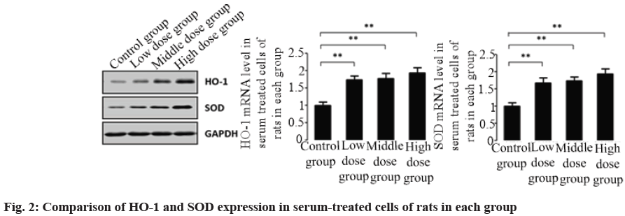
Compared with the control group, the protein levels of HO-1 and SOD in serum-treated cells of rats in Achyranthes bidentata and Radix Paeoniae Alba combined administration group were higher and the mRNA contents of HO-1 and SOD also significantly increased according to qPCR results. Comparison of HO-1 and SOD expression in serum-treated cells of rats in each group is shown as fig. 2.
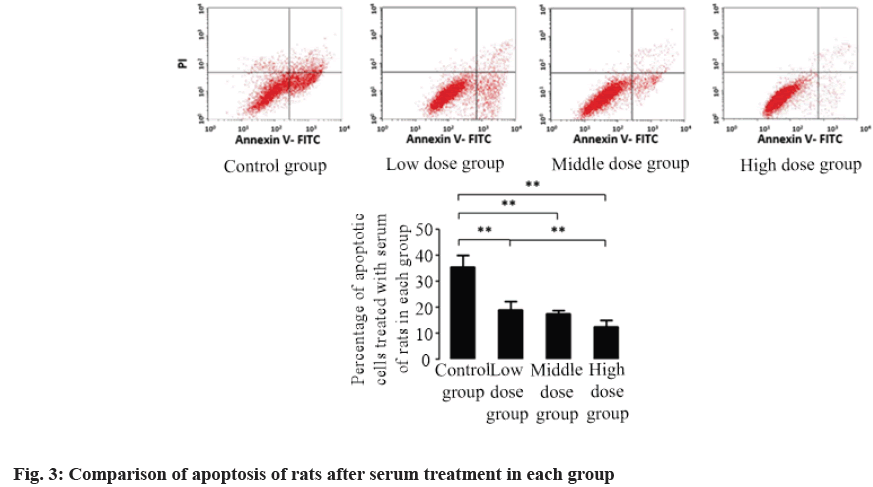
The apoptosis of serum-treated cells in each group was detected by flow cytometry. The results showed that compared with the control group, the proportion of apoptosis in serum-treated cells in Achyranthes bidentata and Radix Paeoniae Alba group was lower (p<0.05). Compared with the low dose group, the apoptosis rate of rats after serum treatment in high dose group was lower (p<0.05). Comparison of apoptosis of rats after serum treatment in each group is shown in fig. 3.
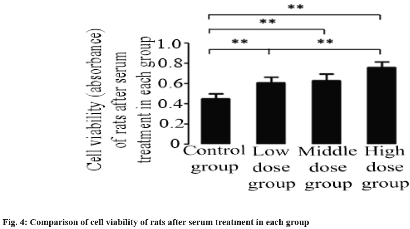
The MTT test showed that compared with the control group, the cell activity of rats after serum treatment in Achyranthes bidentata and Radix Paeoniae Alba combined administration group was higher (p<0.05). Compared with the low dose group, the high dose group showed higher cell activity after serum treatment (p<0.05). Comparison of cell viability of rats after serum treatment in each group is shown in fig. 4.
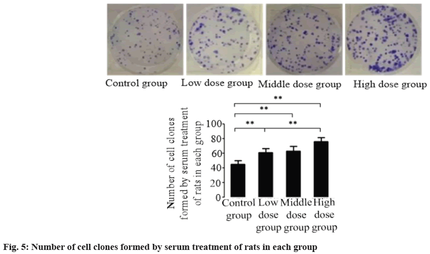
Clone formation test was carried out on the cells of rats after serum treatment in each group. According to the results, compared with the control group, the cells of rats after serum treatment in Achyranthes bidentata and Radix Paeoniae Alba combined administration group had stronger viability (p<0.05). Number of cell clones formed by serum treatment of rats in each group is shown in fig. 5.
Oxidative damage is a process of unbalanced generation and elimination of free radicals in the body or cells, which leads to the accumulation of Reactive Oxygen Species (ROS) and active nitrogen, and finally causes damage. The results of this paper first confirmed that the compatibility of Achyranthes bidentata and Radix Paeoniae Alba could reduce the level of MDA in rat serum, increase the activity of SOD and have the function of resisting oxidative damage. We treated the cells with drug-containing rat serum to simulate the drug action and explored the molecular mechanism of resisting oxidative damage caused by the compatibility of Achyranthes bidentata and Radix Paeoniae Alba. The results showed that two important signal pathways PKCδ and Akt were activated and acted on Nrf2 together, which finally promoted the expression of anti-oxidative enzymes such as NO-1 and SOD, and achieved resistance of oxidative stress damage.
Nrf2 is the key regulator to maintain redox balance. PI3K/AKT pathway can induce the transcription of downstream target gene Nrf2 and then phosphorylate it into the nucleus to combine with Antioxidant Response Element (ARE), thus exerting the cell protection function. Nrf2 which is not phosphorylated into the nucleus is ubiquitinated and degraded in cytoplasm. Previous studies have shown that Oxidized Low Density Lipoprotein (oxLDL) can nitrosate p85 subunit of PI3K, significantly reduce the ratio of Phosphorylated AKT (p-AKT)/AKT and then induce apoptosis. In vitro antioxidant therapy can reverse this phenomenon, which suggests that oxidative stress can interfere with PI3K/ AKT signal transduction process[12,13]. In this study, the effects of Achyranthes bidentata and Radix Paeoniae Alba on Akt pathway and Nrf2 expression were examined. The results showed that the phosphorylation ratio of Akt increased, which promoted the expression of Nrf2. Meanwhile, Nrf2/ARE pathway protected the normal signal transduction of PI3K/AKT pathway through anti-oxidative stress.
It has been reported that the formation and development of osteoarthritis is closely related to oxidative stress[14]. Mechanical stress, aging and other factors of osteoarthritis participate in the development of the disease by improving the level of oxidative stress in the body. Excessive superoxide produced by stress not only leads to mitochondrial dysfunction of chondrocytes, but also promotes chondrocyte apoptosis, synovial cell death and even subchondral ossification of joints. Achyranthes bidentata and Radix Paeoniae Alba pall are often used to treat osteoarthritis and other diseases. This study preliminarily reveals the mechanism of anti-oxidation, anti-inflammation and anti-apoptosis of Achyranthes bidentata pall. It provides a theoretical basis for its clinical application.
Recent studies have found that the overexpression of Nrf2 has carcinogenic effect, but the molecular mechanism of the carcinogenic effect of excessive Nrf2 is still not fully understood, which needs further study[15]. The PI3K/AKT and PKCδ signal pathways involved in this study promote each other and form positive feedback regulation with Nrf2. How to better control the expression level of Nrf2 has become a new problem to be studied. The related research shows that endogenous proteins such as BTB Domain and CNC Homolog 1 (Bach1), Tumor protein (p53) and Activating Transcription Factor 3 (ATF3), and exogenous inhibitors such as All-Trans-Retinoic Acid (ATRA), ochratoxin-A, luteolin, crotonol, procyanidins, apigenin and trigonelline can effectively inhibit the activation of Nrf2-ARE signal pathway[4]. It is believed that in the process of continuous exploration, we can discover new indications of Achyranthes bidentata and Radix Paeoniae Alba, and reduce possible side effects by combined use of other drugs, so that traditional Chinese medicine can be rejuvenated.
Acknowledgements:
The research is supported by Excellent Innovation Team Project of Basic Scientific Research Business Cost in Heilongjiang Province, (No. 2019-KYYWF-1276).
Conflict of interests:
The authors declared no conflict of interest.
References
- Harraan D. Aging: A theory based on free radical and radiation chemistry. J Gerontol 1956;11(3):298-300.
[Crossref] [Google Scholar] [PubMed]
- Sies H, Cadenas E. Oxidative stress: Damage to intact cells and organs. Philos Trans R Soc Lond B Biol Sci 1985;311(1152):617-31.
[Crossref] [Google Scholar] [PubMed]
- Shen YH, Chen CX. Research progress of anti-oxidative stress. Chin Pat Med 2019;41(11):2715-9.
- Wang N, Ma H, Qi X. The Research Progress of Nrf2-ARE signaling pathways in the protection of body oxidative stress injury. Med Pharm J Chin People Lib Army 2015;12:21-7.
[Crossref] [Google Scholar] [PubMed]
- Zhao J, Yuan RH, Wang HF. The activation of PI3K/Akt/Nrf2/HO-1 pathway by Huoxue capsule has an antioxidant effect on vascular endothelial cells. J Yunnan Univ Tradit Chin Med 2019;42(3):10-6.
- Shinjo T, Tanaka T, Okuda H, Kawaguchi AT, Oh-Hashi K, Terada Y, et al. Propofol induces nuclear localization of Nrf2 under conditions of oxidative stress in cardiac H9c2 cells. PLoS One 2018;13(4):e0196191.
[Crossref] [Google Scholar] [PubMed]
- Ling L, Tong J, Zeng L. Paeoniflorin improves acute lung injury in sepsis by activating Nrf2/Keap1 signaling pathway. J Sichuan Univ Med Sci 2020;51(5):84-9.
[Crossref] [Google Scholar] [PubMed]
- Liu Y, Wang LN, Cui LL, Zhang XJ, Ji H. Artesunate improved antioxidant defences and reduced brain damage by activating Nrf2 pathway in a mouse stroke model. J Brain Nerv Dis 2021;29(6):331-5.
- Ma ZY, Zhang DI, Sun J, Niu J, Ren Q, Wu QW, et al. Forsythiaside A inhibited cerebral ischemic induced oxidative damage through Akt/Nrf2 signaling pathway. Prog Mod Biomed 2021;21(2):214-8.
- Yang YJ, Yang GY, Yu HS, Guan XF. Analysis on ?Component-target-pathway? of Achyranthes bidentata Blume and Paeonia lactiflora Pall drug pair in treating osteoarthritis based on mining and integrated pharmacology. J Liaoning Univ Tradit Chin Med 2020;22(3):127-31.
- Li WX, Wang WH. Experimental study on the treatment of lumbar disc herniation with Duhuo Jisheng decoction with different processed products. Strait Pharm J 2021;33(6):12-5.
- Tie G, Yan J, Yang Y, Park BD, Messina JA, Raffai RL, et al. Oxidized low-density lipoprotein induces apoptosis in endothelial progenitor cells by inactivating the phosphoinositide 3-kinase/Akt pathway. J Vasc Res 2010;47(6):519-30.
[Crossref] [Google Scholar] [PubMed]
- Li C, Xia JY, Wan Q. Effect of interaction between Nrf2/ARE pathway and PI3K/AKT pathway on caspase -3 expression in islet cells. Chongqing Med J 2017;46(3):292-5.
- Xia GQ. Research progress on the correlation between oxidative stress and osteoarthritis and the application of anti-oxidative stress drugs. Chin J Diffic Complicated Cases 2021;20(1):94-8.
-
Namani A, Li Y, Wang XJ, Tang X. Modulation of NRF2 signaling pathway by nuclear receptors: Implications for cancer. Biochim Biophys Acta Mol Cell Res 2014;1843(9):1875-85.
[Crossref] [Google Scholar] [PubMed]


