- *Corresponding Author:
- Dongqing Xu
Department of Health and Exercise Science, Tianjin University of Sports, Hexi, Tianjin 300060, China
E-mail: xudongqing54@163.com
| This article was originally published in a special issue, “Role of Biomedicine in Pharmaceutical Sciences” |
| Indian J Pharm Sci 2023:85(2) Spl Issue “96-102” |
This is an open access article distributed under the terms of the Creative Commons Attribution-NonCommercial-ShareAlike 3.0 License, which allows others to remix, tweak, and build upon the work non-commercially, as long as the author is credited and the new creations are licensed under the identical terms
Abstract
Knee osteoarthritis is the most common degenerative bone and joint disease in the middle-aged and elderly. Here, we compared the diverse formulations of sports intensity and pharmaceutical selection for knee osteoarthritis patients. In this study, we compared and analyzed the differences and characteristics of lower limb joint kinematics, dynamics and muscle contraction function of knee osteoarthritis patients when walking at different speeds and the ones taking pharmaceutical selection including oral non-steroidal antiinflammatory drugs as well as topical capsaicin, so as to explore the impact of walking speed and drugs on the walking ability of knee osteoarthritis patients. 16 patients with left knee osteoarthritis and 16 patients with right knee osteoarthritis were selected and were grouped for different walking speed; 8 subjects were given oral non-steroidal anti-inflammatory drugs and 8 subjects were given topical capsaicin. Kistler threedimensional dynamometer, Noraxon surface electromyography and Qualisys infrared three-dimensional motion analysis were used to record the gait parameters. Compared with walking at low speed, normal walking speed significantly increased the knee maximum flexion angle. For dynamic parameters, the lowest value of first peak knee adduction moment presented in normal speed patients and there was also significant difference between groups for hip flexion torque (p=0.014), the first peak of knee adduction torque (p=0.043) and knee flexion torque (p=0.040). For the electromyographic parameters, in the early period of support, the speed of the lower extremity rectus femoris (p=0.001), anterior tibial muscle (p=0.016), lateral femur muscle (p=0.040) and the average amplitude of electromyography were not equal for distinct groups. We found that the outcome of kinematics parameters for oral non-steroidal anti-inflammatory drug was greatly higher than topical capsaicin. The research conclusion will provide important scientific basis and theoretical support for knee osteoarthritis population to conduct walking exercise scientifically and reasonably.
Keywords
Walking speed, knee osteoarthritis, nordic walking, non-steroidal anti-inflammatory drugs, topical capsaicin
With the deepening trend of social aging, more and more senile diseases need to be paid attention. Knee Osteoarthritis (KOA) is the most common degenerative bone and joint disease in the middleaged and elderly[1]. Due to joint pain, swelling, deformity and other reasons, KOA patients have limited mobility, which not only seriously affects their quality of life, but also leads to a significant increase in the incidence of cardiovascular events and all-cause mortality[2,3]. Therefore, the prevention and treatment of KOA is particularly important.
Exercise therapy is an active means to prevent and treat KOA[4]. By increasing muscle strength, improving endurance, improving and maintaining joint function, exercise can effectively delay the progress of disease and improve the quality of life of patients[5]. Although the exercise training for KOA patients should be particularly careful in terms of intensity, formulation selection, etc., which still needs more research and demonstration. Exercise therapy has been recommended as one of the basic treatment methods in the guidelines for the diagnosis and treatment of osteoarthritis at home and abroad[6]. Walking exercise is a common form of exercise for KOA patients[7], which is convenient and easy to control exercise intensity. However, due to the destruction of articular cartilage and the decline of lower limb muscle function and other obstacles, changes in exercise intensity and exercise volume during walking of KOA patients are very likely to cause problems such as excessive joint load and abnormal stress distribution, thus increasing the risk of disease deterioration[8]. Walking speed not only determines the intensity of walking exercise, affects the amount of exercise, but also is an important factor to change the load of lower limb joints. The change of walking speed can be the active adjustment of the human body to cope with different needs or it can be the passive compensation when the body structure is damaged. Therefore, walking speed is also one of the important indicators reflecting walking ability. Many studies have shown that the walking speed of KOA patients decreases, which is considered as a compensatory reaction to reduce joint load[9].
Except for physical approaches, a variety of pharmacologic agents have been also recommended for KOA patients, including oral Non-Steroidal Anti- Inflammatory Drugs (NSAIDs) and topical capsaicin. A large number of trials have established their shortterm efficacy[10].
Based on the fact of rapid increasing population of KOA patients, the best treatment approach is uncertain yet. Therefore, this study shows the impact of the KOA population of various key factors, to compare and analyze the differences between diverse formulations of sports intensity and pharmaceutical selection. The conclusion of this study will provide scientific basis and theoretical support for KOA people to walk scientifically and rationally.
Materials and Methods
Subjects:
32 KOA patients with chronic KOA were recruited through various sources (medical site recruitment, media publicity, health lectures, etc.). According to the following standards, all were unilateral patients, 16 patients with left KOA and 16 patients with right KOA, aged 55-65 y old[11].
Screening criteria: Subjects aged>55 y were screened according to the KOA diagnostic criteria of United States College of Rheumatology and patients with Kellgren-Lawrence (KL) grade II or III. If that patient did not take analgesics within 1 mo[12,13], the same orthopedic surgeon should carry out the corresponding clinical grading according to the medical record of the third-class hospital presented by the patient, in combination with the X-ray film taken by the hospital when seeing a doctor and the diagnosis report thereof.
Exclusion criteria: Patients with the following conditions were excluded from this study. Patients with secondary KOA; those who need crutches for walking; patients who have been injected analgesics into articular cavity in the past 3 mo; hypertension and hyperglycemia are difficult to control; patients with lumbar diseases and plantar diseases; patients who had knee or hip surgery or fracture of lower limb in the past 3 mo; obese (Body Mass Index (BMI)>28 kg/m2) and allergy to oral NSAID and topical capsaicin. Basic information of subjects was shown in Table 1.
| Sample size | Age (y) | Height (cm) | Weight (kg) | BMI (kg/m2) |
|---|---|---|---|---|
| 32 (male: 14; female: 18) | 60.33±3.65 | 165.3±6.58 | 66.26±8.04 | 24.24±3.41 |
Table 1: Basic Information of Subjects
Administration of pharmacologic agents:
16 subjects were grouped for different walking speed. 8 subjects were given oral NSAID (celecoxib, 100 mg twice a day, Pfizer Inc.) and 8 subjects were given topical capsaicin (100 mg twice a day, Pfizer Inc.), respectively.
Experimental methods:
Kistler three-Dimensional (3D) dynamometer, Noraxon surface Electromyography (EMG) and Qualisys infrared three-dimensional motion analysis were used to record the gait parameters of KOA patients during walking at different speeds [14].
Morphological index measurement:
According to the testing method in practical physique, the same experimenter adopts the height and weight measuring instrument in the national physique monitoring instrument to collect the data of subject’s height and weight.
Gait test:
The experiment was carried out in the biomechanics laboratory of the university[15,16]. The subjects were tested with walking speed at 8×1.2 meters. The Kistler 3D dynamometer was positioned in the middle of the channel. The Qualisys OQUS300+ series optical motion capture system (8 cameras) was placed in 8 corners, allowing it to cover the entire walkway. Qualisys with a synchronous interface, the force platform, surface EMG and 3D infrared motion capture system for synchronous acquisition.
The normal speed (1.02±0.08 m/s) is the comfortable walking speed selected by the subject and the slow speed (0.90±0.07 m/s) and fast speed (1.14±0.09 m/s) are 15 % slower and faster than their normal walking speed respectively. The subjects without drug treatment undertook different speeds while the subjects with drug treatment undertook normal speed.
Statistical analysis:
Statistical Package for the Social Sciences (SPSS) 22.0 software was used to analyze and test the obtained experimental data. The differences of each test index of the two walking forms were tested and compared by the paired sample t-test and the multivariate variances were measured repeatedly to analyze the discrepancies of each test index under the three-phase asynchronous velocities. The significance level was determined as p<0.05.
Results and Discussion
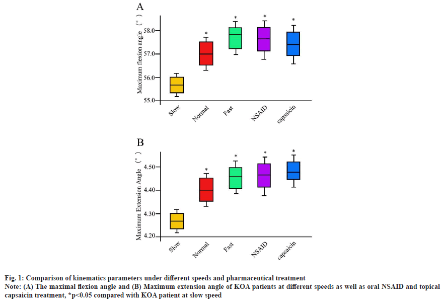
Comparison of kinematics parameters under different speeds and pharmaceutical treatment was shown in fig. 1. Compared with walking at low speed, normal walking speed significantly increased the knee maximum flexion angle. The high speed walking exhibited the maximal flexion angle, which was similar to NSAID treatment (fig. 1A). The topical capsaicin treatment was relatively lower for knee maximum flexion angle. On the other hand, the maximum extension angle of patients with pharmaceutical treatment was significantly greater than that with exercises and the low speed exercise patients were lowest in first peak knee adduction moment (fig. 1B).
Fig. 1: Comparison of kinematics parameters under different speeds and pharmaceutical treatment
Note: (A) The maximal flexion angle and (B) Maximum extension angle of KOA patients at different speeds as well as oral NSAID and topical
capsaicin treatment, *p<0.05 compared with KOA patient at slow speed
There were also remarkable differences for other kinematics parameters between distinct groups i.e. walking frequency (p=0.000), step length (p=0.026), duration of bipedal support (p=0.013) and total duration of support (p=0.000).
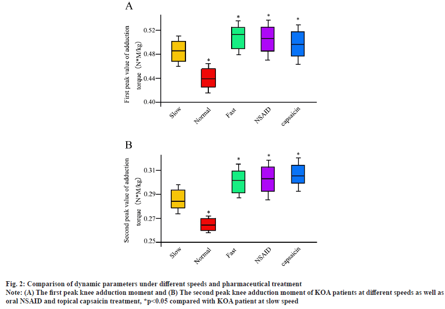
Comparison of dynamic parameters under different speeds and pharmaceutical treatment was shown in fig. 2A and fig. 2B. For dynamic parameters, the lowest value of first peak knee adduction moment is presented in normal speed patients. While, both slow and fast speed enhanced the first peak knee adduction moment significantly. At the same time, both NSAID and topical capsaicin treatment increased the results of first peak knee adduction moment. The second peak knee adduction moment demonstrated the similar pattern with the first peak knee adduction moment and there was also significant difference between groups for hip flexion torque (p=0.014), the first peak of knee adduction torque (p=0.043) and knee flexion torque (p=0.040).
Fig. 2: Comparison of dynamic parameters under different speeds and pharmaceutical treatment
Note: (A) The first peak knee adduction moment and (B) The second peak knee adduction moment of KOA patients at different speeds as well as
oral NSAID and topical capsaicin treatment, *p<0.05 compared with KOA patient at slow speed
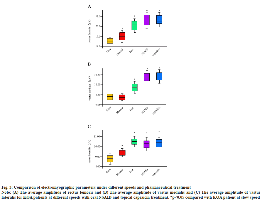
Comparison of electromyographic parameters under different speeds and pharmaceutical treatment was shown in fig. 3A-fig. 3C. For the electromyographic parameters, in the early period of support, the speed of the lower extremity rectus femoris (p=0.001), anterior tibial muscle (p=0.016), lateral femur muscle (p=0.040) and the average amplitude of EMG were not equal for distinct groups. To be specific, the average amplitude of the rectus femoris muscle in the fast walking was greater than that in the normal walking; the average amplitude of the anterior tibial muscle in the normal walking was greater than that in the slow walking. Both NSAID and topical capsaicin treatment increased the results of amplitude of the rectus femoris. At the same time, there are remarkable differences for the lateral femoris muscle among distinct groups. The overall tendency of amplitude of vastus medialis was similar to rectus femoris. However, there was no significant difference between slow speed and normal speed. There was no evident difference in the mean amplitude of EMG between normal speed and slow speed.
Fig. 3: Comparison of electromyographic parameters under different speeds and pharmaceutical treatment
Note: (A) The average amplitude of rectus femoris and (B) The average amplitude of vastus medialis and (C) The average amplitude of vastus
lateralis for KOA patients at different speeds with oral NSAID and topical capsaicin treatment, *p<0.05 compared with KOA patient at slow speed
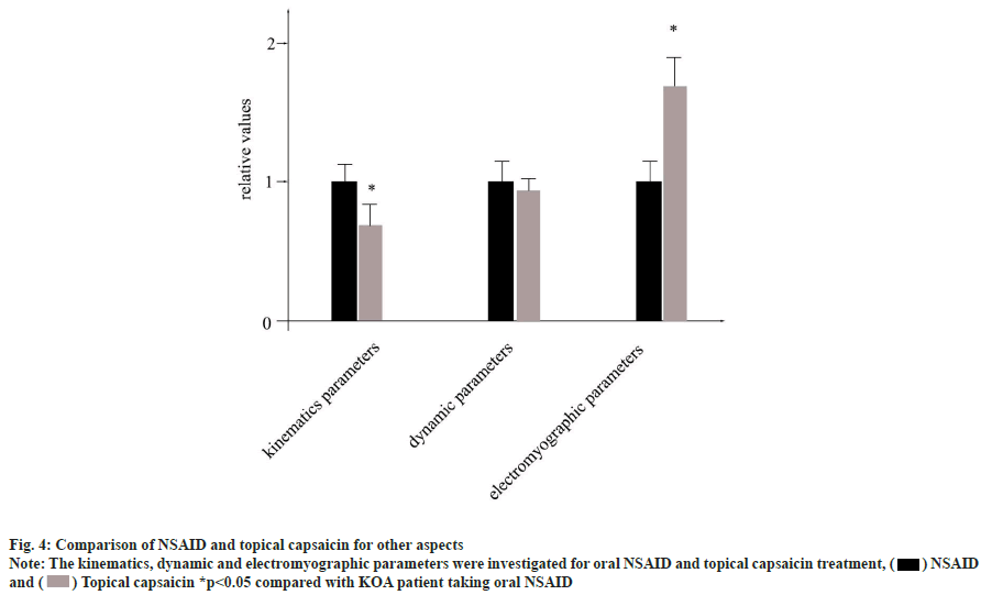
Comparison of NSAID and topical capsaicin for other aspects was shown in fig. 4. Both oral NSAID and topical capsaicin are primary recommended approaches for KOA patients. However, still there is lack of systematical comparison of them for the disorder yet. To this end, here we sought to compare them from three aspects (kinematics parameters, dynamic parameters and electromyographic parameters) in detail. The flexion extension range was considered as a representative for kinematics parameters, abduction moment was considered as a representative for dynamic parameters and the average amplitude of anterior tibial muscle was considered as a representative for electromyographic parameters.
As shown in fig. 4, the outcome of kinematics parameters for oral NSAID was greatly higher than topical capsaicin. Inversely, the results of electromyographic parameters in KOA patients taking topical capsaicin were significantly greater than KOA patients using oral NSAID. There was no remarkable difference for dynamic parameters between two groups of KOA patients.
Based on the results of this study, we can find that the walking speed is an important factor affecting the movement of lower limb joints and muscles in KOA patients. However, the influence of different speeds on related gait parameters is not the same. Compared with walking at slow speed, KOA patients have better adaptability to the change of walking speed and they did a better performance at faster speed for kinematics parameters and electromyographic parameters, which was consistent with previously reported[17-19]. However, fast speed is beneficial for KOA patients at all three respects including kinematics parameters, dynamic parameters as well as electromyographic parameters, Moreover, in all the three entries, the fast speed training exercise is comparable with oral NSAID and topical capsaicin administration, suggesting an efficient physical replacement for medical approach.
In normal walking, KOA patients mainly improve walking speed by increasing walking frequency and shortening supporting time[20,21]. Compared with the normal speed, the first peak of knee adduction torque in the strut period did not increase when walking at a fast pace 15 % higher than normal speed, but it may be necessary to compensate the lower limb load by increasing the abduction moment of hip and knee. Although some muscle activities of lower limbs are enhanced during rapid general walking, the impact on lower limb load should be considered in order to increase the walking speed and increase the exercise intensity. When walking at a pace 15 % lower than the normal walking speed, the lower limb torque of KOA patients reduced, which has the advantage of reducing the load on the lower limb and the degree of muscle contraction is not weakened by the reduction of the walking speed. The reduction of speed may not be conducive to the improvement of physical activity level in patients with KOA.
When we observed the KOA patient with oral NSAID and topical capsaicin treatment, they clearly exhibited overall outcomes similar to the patient with increased step frequency, increased step length and shortened supporting time, which is closer to the changes in the temporal and spatial parameters of the gait of normal people. Compared with walking with physical training, pharmacologic method can mobilize more muscle activities and have more obvious advantages for muscle training. For them, the first peak of adductive torque is significantly increased and the ranges of hip and knee extension reduced, the torque decreased and the degree of muscle contraction did not change too much. It has the advantage of better reducing the load of lower limbs without affecting the lower limbs muscles, which is similar to previously reported[22-24].
Even popularly used in clinic, there is still missing an in-depth comparison of oral NSAID and topical capsaicin for KOA patients. Here, we could show that oral NSAID and topical capsaicin were not equally functional for KOA patients. The treatment of oral NSAID played an important role in kinematics parameters but not in electromyographic parameters for KOA patient while, both chemical drugs were equally useful for dynamic parameters of KOA patients. When we looked at the results in detail, we found that the first peak of adductive moment of the knee joint during the topical capsaicin was lower than that of oral NSAID and the topical capsaicin had less impact on the lower limb load of KOA patients.
All in all, these might suggested that oral NSAID approach not only fitted the needs of KOA patients, but also had a greater tolerance to adjust the exercise intensity. Overall, our study here systematically compared the advantages and disadvantages of physical exercises and distinct pharmacologic treatments. All the results here provided several beneficial hints for future clinical selections of KOA patients.
Conflict of interests:
The authors declared no conflict of interest.
References
- Tang X, Wang S, Zhan S, Niu J, Tao K, Zhang Y, et al. The prevalence of symptomatic knee osteoarthritis in China: Results from the China health and retirement longitudinal study. Arthritis Rheumatol 2016;68(3):648-53.
[Crossref] [Google Scholar] [PubMed]
- Hawker GA, Croxford R, Bierman AS, Harvey PJ, Ravi B, Stanaitis I, et al. All-cause mortality and serious cardiovascular events in people with hip and knee osteoarthritis: A population based cohort study. PLoS One 2014;9(3):e91286.
[Crossref] [Google Scholar] [PubMed]
- McAlindon TE, Bannuru R, Sullivan MC, Arden NK, Berenbaum F, Bierma-Zeinstra SM, et al. OARSI guidelines for the non-surgical management of knee osteoarthritis. Osteoarthritis Cartilage 2014;22(3):363-88.
[Crossref] [Google Scholar] [PubMed]
- Hootman JM, Macera CA, Ham SA, Helmick CG, Sniezek JE. Physical activity levels among the general US adult population and in adults with and without arthritis. Arthritis Rheum 2003;49(1):129-35.
[Crossref] [Google Scholar] [PubMed]
- Kovar PA, Allegrante JP, MacKenzie CR, Peterson MG, Gutin B, Charlson ME. Supervised fitness walking in patients with osteoarthritis of the knee: A randomized, controlled trial. Ann Intern Med 1992;116(7):529-34.
[Crossref] [Google Scholar] [PubMed]
- Farrokhi S, Jayabalan P, Gustafson JA, Klatt BA, Sowa GA, Piva SR. The influence of continuous versus interval walking exercise on knee joint loading and pain in patients with knee osteoarthritis. Gait Posture 2017;56:129-33.
[Crossref] [Google Scholar] [PubMed]
- Roddy E, Zhang W, Doherty M. Aerobic walking or strengthening exercise for osteoarthritis of the knee? A systematic review. Ann Rheum Dis 2005;64(4):544-8.
[Crossref] [Google Scholar] [PubMed]
- White DK, Tudor-Locke C, Felson DT, Gross KD, Niu J, Nevitt M, et al. Walking to meet physical activity guidelines in knee osteoarthritis: Is 10,000 steps enough? Arch Phys Med Rehabil 2013;94(4):711-7.
[Crossref] [Google Scholar] [PubMed]
- Messier SP, Loeser RF, Miller GD, Morgan TM, Rejeski WJ, Sevick MA, et al. Exercise and dietary weight loss in overweight and obese older adults with knee osteoarthritis: The arthritis, diet, and activity promotion trial. Arthritis Rheum 2004;50(5):1501-10.
[Crossref] [Google Scholar] [PubMed]
- Wallis JA, Webster KE, Levinger P, Singh PJ, Fong C, Taylor NF. The maximum tolerated dose of walking for people with severe osteoarthritis of the knee: A phase I trial. Osteoarthritis Cartilage 2015;23(8):1285-93.
[Crossref] [Google Scholar] [PubMed]
- Mündermann A, Dyrby CO, Hurwitz DE, Sharma L, Andriacchi TP. Potential strategies to reduce medial compartment loading in patients with knee osteoarthritis of varying severity: Reduced walking speed. Arthritis Rheum 2004;50(4):1172-8.
- Fernandes WC, Machado Á, Borella C, Carpes FP. Influence of gait speed on plantar pressure in subjects with unilateral knee osteoarthritis. Rev Bras Reumatol 2014;54:441-5.
[Crossref] [Google Scholar] [PubMed]
- Schiffer T, Knicker A, Hoffman U, Harwig B, Hollmann W, Strüder HK. Physiological responses to nordic walking, walking and jogging. Eur J Appl Physiol 2006;98(1):56-61.
[Crossref] [Google Scholar] [PubMed]
- Chan GN, Smith AW, Kirtley C, Tsang WW. Changes in knee moments with contralateral versus ipsilateral cane usage in females with knee osteoarthritis. Clin Biomech 2005;20(4):396-404.
[Crossref] [Google Scholar] [PubMed]
- Bohne M, Abendroth-Smith J. Effects of hiking downhill using trekking poles while carrying external loads. Med Sci Sports Exerc 2007;39(1):177-83.
[Crossref] [Google Scholar] [PubMed]
- Schwameder H, Ring S. Knee joint loading and metabolic energy demand in walking, nordic walking and running. J Biomech 2006(39):S185.
- Stief F, Kleindienst FI, Wiemeyer J, Wedel F, Campe S, Krabbe B. Inverse dynamic analysis of the lower extremities during nordic walking, walking, and running. J Appl Biomech 2008;24(4):351-9.
[Crossref] [Google Scholar] [PubMed]
- Hagen M, Hennig EM, Stieldorf P. Lower and upper extremity loading in nordic walking in comparison with walking and running. J Appl Biomech 2011;27(1):22-31.
[Crossref] [Google Scholar] [PubMed]
- Zhu W. Let's keep walking. Med Sci Sports Exerc 2008;40(7):S509-11.
[Crossref] [Google Scholar] [PubMed]
- Robertson MC, Campbell AJ, Gardner MM, Devlin N. Preventing injuries in older people by preventing falls: A meta-analysis of individual-level data. J Am Geriatr Soc 2002;50(5):905-11.
[Crossref] [Google Scholar] [PubMed]
- Ebrahim S, Thompson PW, Baskaran V, Evans K. Randomized placebo-controlled trial of brisk walking in the prevention of postmenopausal osteoporosis. Age Ageing 1997;26(4):253-60.
[Crossref] [Google Scholar] [PubMed]
- Nelson ME, Fisher EC, Dilmanian FA, Dallal GE, Evans WJ. A 1-y walking program and increased dietary calcium in postmenopausal women: Effects on bone. The Am J Clin Nutr 1991;53(5):1304-11.
[Crossref] [Google Scholar] [PubMed]
- Kohrt WM, Ehsani AA, Birge JR SJ. Effects of exercise involving predominantly either joint-reaction or ground-reaction forces on bone mineral density in older women. J Bone Miner Res 1997;12(8):1253-61.
[Crossref] [Google Scholar] [PubMed]
- Devos-Comby L, Cronan T, Roesch SC. Do exercise and self-management interventions benefit patients with osteoarthritis of the knee? A metaanalytic review. J Rheumatol 2006; 33(4):744-56.
[Google Scholar] [PubMed]


 ) NSAID
and (
 ) Topical capsaicin *p<0.05 compared with KOA patient taking oral NSAID


