- *Corresponding Author:
- Yiling Hou
Key Laboratory of Southwest China Wildlife Resources Conservation (Ministry of Education), College of Life Sciences, China West Normal University, Nanchong, 637009, China
E-mail: starthlh@126.com
| Date of Submission | 22 April 2017 |
| Date of Revision | 23 March 2018 |
| Date of Acceptance | 22 September 2018 |
| Indian J Pharm Sci 2018;80(6):1029-1038 |
This is an open access article distributed under the terms of the Creative Commons Attribution-NonCommercial-ShareAlike 3.0 License, which allows others to remix, tweak, and build upon the work non-commercially, as long as the author is credited and the new creations are licensed under the identical terms
Abstract
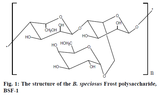
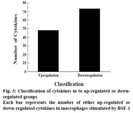
The polysaccharide of Boletus speciosus Frost has a backbone of (1→4)-α-L-mannopyranose residues, which branched at O-6 and the branches mainly comprised of (a→1)-α-D-galactopyranose residue. To find out signaling transduction pathways that mediate the effect of the polysaccharide of Boletus speciosus Frost on macrophages, the polysaccharide of B. speciosus Frost was used to stimulate the macrophages and 200 cytokines were detected with the help of a protein chip. Further analysis of the result revealed 48 up-regulated and 73 down-regulated cytokines. All cytokines were imported into the KEGG Pathway Database and NCBI to identify the signaling pathway and biological functions involved, respectively. The results showed that the cytokines fulfilled valuable functions in the JAK-STAT signaling pathway and NF-kappa B signaling pathway. This study thus revealed that the edible fungus polysaccharides could stimulate immune cells to secrete immune factors, which could provide a scientific basis for targeting sugar immunization preparations.
Keywords
Boletus speciosus Frost, polysaccharide, protein chip, macrophages
Scientific experimental studies showed that many edible fungus polysaccharides had biological activities, including immune regulation and antitumor activities [1]. It is becoming increasingly clear by using the technology in modern medicine, cell biology and molecular biology to know how the immune system works. It would lead to the occurrence of human aging and a variety of diseases if the immune system is disordered [2]. Immune regulation by polysaccharides plays a role through the activation of macrophages, T and B lymphocytes, the reticuloendothelial system, complement and interferon, interleukin (IL) generation [3]. In addition, scientists further found that macrophages are one of the most important immune cells. It was and remained to be an integral part in studying cell phagocytosis, cellular immunity and molecular immunology [4]. With the stimulation of antigen, innate immune response is produced, which is not only sterilize directly but also secrete innate immune factors such as cell factors and chemotactic factors to regulate the function of the immune system [5]. Boletus speciosus Frost, a very rare edible fungus with high nutritional value, also has important medicinal values such as promoting digestion and enhancing immunity. Some progress has been made on bioactive substances from B. speciosus, for example some important components of B. speciosus were responsible for anticancer [6], antioxidant activities and for lowering blood lipids. In our previous studies, the polysaccharide structure of B. speciosus, which was named BSF-1 had been identified. The molecular weight of BSF-1 was 1.33×10⁴ Da. BSF-1 consisted of two kinds of monosaccharides which were L-Man and D-Gal in the ratios of 2:1. It had a backbone of (1→4)-α-L-mannopyranose residues, which branches at O-6. The branches mainly comprised of (a→1)-α-Dgalactopyranose residue (Figure 1) [7]. A protein chip was used to identify the signalling transduction pathways in macrophages stimulated by polysaccharide BSF-1.
Materials and Methods
The fruiting bodies of B. speciosus were collected in Sichuan province, China and were authenticated in the College of Life Sciences, China West Normal University, Nanchong, China. DEAE-cellulose was purchased from Sigma-Aldrich (Mainland, China). Monosaccharide standards were purchased from Beijing BioDee Biotechnology Co. Ltd. (Beijing, China). All other reagents used were of analytical grade.
Extraction and purification of polysaccharides from B. speciosus Frost
In order to remove lipids, B. speciosus Frost fruiting bodies were soaked with 95 % alcohol for 6 h. The powder of B. speciosus Frost fruiting bodies was boiled three times each time for 6 h to extract polysaccharides. The filtrate was concentrated, dialyzed (MWCO8000, Sigma) and centrifuged to remove insoluble material and small molecular compounds [8]. Three times volume of 95 % alcohol was added to precipitate the crude polysaccharides, which was dried at 65°. Polysaccharides of B. speciosus Frost were extracted and purified using DEAE-Sepharose fast flow column [9].
Cell culture experiments
After macrophages (RAW 264.7 cells, derived from mice, Chengdu golden Kay Biological Technology Co. Ltd. Chengdu, China) were recovered, 1640 basic culture medium (phenol red free) was used to culture the macrophages. The composition of the culture medium was as follows, 90 % RPMI-1640 culture medium (phenol red free), 10 % fetal bovine serum and 1 % double antibiotics (10 mg/ml streptomycin and 10000 U/ml penicillin) [10]. Macrophages were digested using 0.5 % pancreatin when cells were in the logarithmic growth phase. The cell culture fluid was added to dilute macrophages at a density of 2×10⁵ cells/ml.
The polysaccharide BSF-1 solution was mixed at concentrations of 2.5, 5, 10 and 20 μg/ml, respectively with 0.2 % fetal bovine serum medium. There were three groups, the blank group, LPS group and the BSF-1 group. Each well was added 1 ml cell dilution solution in the 6-well plates. The 6-well plates were placed in a CO2 incubator (37°) to cultivate. After cultivated for 24 h, the supernatant was removed from the 6-well plates. The blank group of the 6-well plates was added 1 ml 0.2 % fetal bovine serum medium, the LPS group was added 1 ml (10 μg/ml) LPS, and the BSF-1 groups were added 1 ml each of different concentrations of BSF-1 solution (2.5, 5, 10, and 20 μg/ml). The 6-well plates were incubated in 5 % CO2 incubator (37°). After cultivated for 72 h, the supernatant was sucked with a pipette in 2 ml centrifuge tubes and sent to the company (Ray Biotech, Inc., Guangzhou, China) to carry out protein chip experiment.
Statistical analysis
Experimental data was received from the company. Each of the cytokines had data in triplicate for the blank as well as the BSF-1 group. Average and standard deviation of values of each cytokine in the blank and the BSF-1 group were calculated [11]. The level of each cytokine from the BSF-1 group was expressed as a multiple of the corresponding value from the blank group. Student’s t-test and one-way analysis of variance were used for statistical analysis. A level of p<0.05 was considered to be statistically significant. Owing to the abundant experimental data with large changes, logarithm of the original data was used to make the bar graph to make the data easily comprehensible.
Kyoto Encyclopedia of Genes and Genomes (KEGG) enrichment analysis
All the up-regulated or down-regulated cytokines were introduced into KEGG for cell signalling pathway analysis. Pathway Builder Tool 2.0 software was used to draw the map of the identified signalling pathway and NCBI was used to learn about the biological function of the related cytokines and the expression mechanism of protein in macrophage.
Results and Discussion
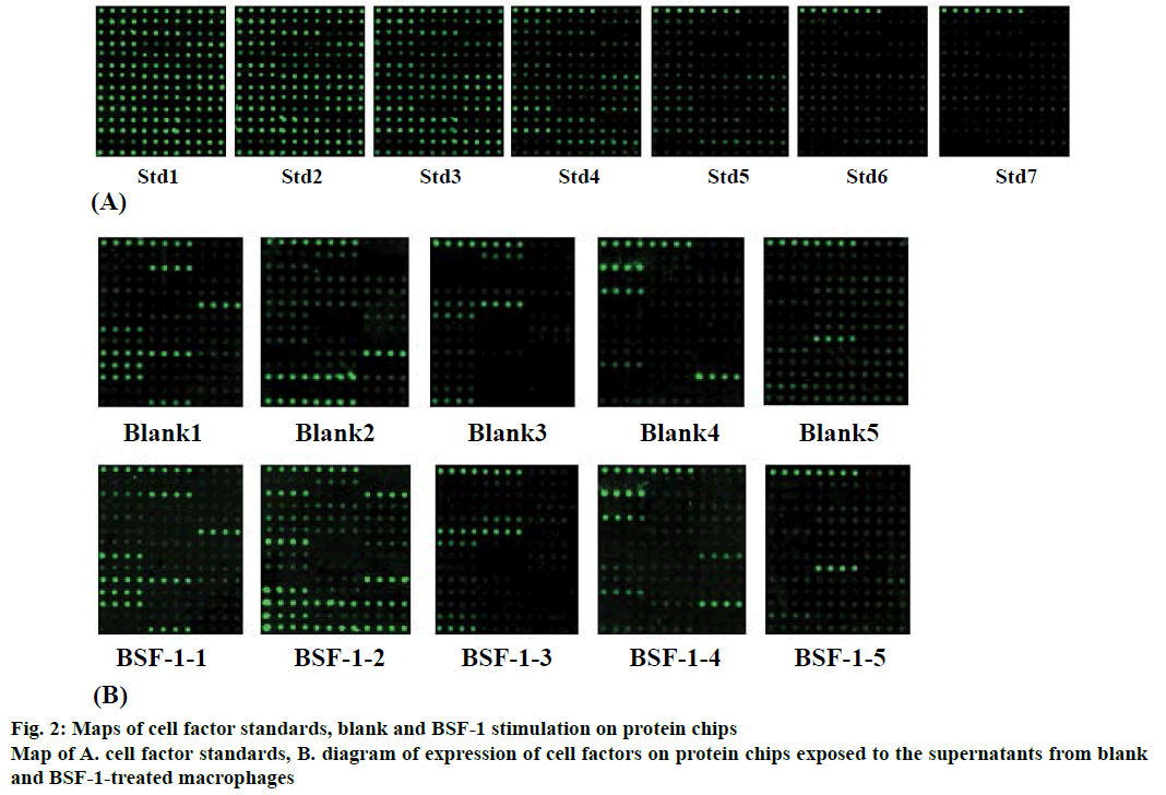
Cells were cultured to collect the supernatant to complete the protein chip experiment. In this experiment, cell factors in macrophages had a standard (Figure 2A). The original diagram of the chip was analyzed (Figure 2B). The results indicated that there were differences in fluorescence intensity between the blank group and BSF-1 group. All the fluorescence intensities were automatically identified by the computer. Two hundred cytokines were tested using five protein chips and the results indicated that there were 48 upregulated cytokines and 73 down-regulated cytokines in macrophages induced by BSF-1 (Figure 3).
Proteins with up-regulated expression meant that certain signalling pathways were activated. That immune systems could operate through these signalling pathways in macrophages was surmised. Through sorting the data, 48 proteins with up-regulated expression were found, such as TNF-α, IL-6, G-CSF, LOX-1 and RANTES. These cytokines were divided into two groups. One group that was 5 to 20 fold up-regulated (Table 1) and the other was 20 fold upregulated (Table 2). Individual cytokines with the number of fold up-regulation in the BSF-1 group relative to the blank group were listed in Tables 1 and 2.
| Cytokines | Blank | BSF-1 | Fold Change | Cytokines | Blank | BSF-1 | Fold Change |
|---|---|---|---|---|---|---|---|
| GITR | 1.1 | 5.7 | 5.0 | Fcg RIIB | 132.5 | 1165.2 | 8.8 |
| PF-4 | 32.6 | 195.2 | 6.0 | I-TAC | 19.3 | 181.1 | 9.4 |
| TSLP | 1.2 | 7.4 | 6.1 | Nope | 4.0 | 37.8 | 9.6 |
| SDF-1α | 30.7 | 199.1 | 6.5 | B7-1 | 40.5 | 397.8 | 9.8 |
| IL-12p40 | 0.0 | 6.5 | 6.5 | P-selectin | 3.3 | 33.1 | 9.9 |
| CD30T | 4.0 | 25.9 | 6.5 | IGFBP-2 | 16.9 | 173.6 | 10.2 |
| VEGF R1 | 18.1 | 119.4 | 6.6 | IGFBP-3 | 31.8 | 331.2 | 10.4 |
| MIP-3β | 0.6 | 3.9 | 6.6 | JAM-A | 0.1 | 1.6 | 11.0 |
| Decorin | 7.8 | 53.3 | 6.8 | MDC | 59.6 | 700.0 | 11.7 |
| IL-20 | 0.0 | 6.9 | 6.9 | Pro-MMP-9 | 2685.2 | 31914.0 | 11.9 |
| CD27L | 1.9 | 13.5 | 7.1 | MIP-3α | 0.3 | 3.6 | 13.7 |
| Eotaxin-2 | 19.7 | 141.0 | 7.2 | IL-12p70 | 0.0 | 14.6 | 14.6 |
| IL-1β | 7.4 | 53.5 | 7.3 | VCAM-1 | 0.0 | 14.9 | 14.9 |
| Fas L | 1.9 | 14.5 | 7.8 | IL-1RA | 270.5 | 4578.0 | 16.9 |
| AR | 3.7 | 32.7 | 8.8 | IL-1a | 19.6 | 361.2 | 18.4 |
Table 1: 5-20 Fold up-Regulated Cytokine Expression in Macrophages Stimulated with BSF-1 Relative to the Blank Group
| Cytokines | Blank | BSF-1 | Fold up-regulation | Cytokines | Blank | BSF-1 | Fold up-regulation |
|---|---|---|---|---|---|---|---|
| TARC | 2.4 | 55.8 | 23.6 | IL-23 | 0.0 | 79.8 | 79.8 |
| IL-28 | 1.2 | 33.7 | 27.8 | NOV | 0.0 | 94.5 | 94.5 |
| Meteorin | 0.0 | 28.7 | 28.7 | MCP-5 | 2.2 | 313.7 | 145.6 |
| CD40 | 18.7 | 581.0 | 31.1 | IL-6 | 95.5 | 25161.1 | 263.5 |
| PlGF-2 | 1.3 | 42.6 | 32.9 | Lipocalin-2 | 14.7 | 11833.1 | 806.2 |
| DAN | 0.0 | 45.4 | 45.4 | TNFα | 14.0 | 15773.7 | 1127.9 |
| Lymphotactin | 0.0 | 46.9 | 46.9 | RANTES | 0.8 | 1312.4 | 1694.7 |
| TREM-1 | 2.7 | 134.2 | 49.7 | LOX-1 | 0.2 | 382.8 | 1925.1 |
| DLL4 | 13.2 | 654.7 | 49.7 | G-CSF | 0.0 | 3766.5 | 3766.5 |
Table 2: More Than 20 Fold up-Regulated Cytokines in Macrophages of BSF-1 Group Relative to the Blank Group
Analysis of the processed data revealed a large IL family with in the 5.0 to 20.0 fold up-regulated group of cytokines that included IL-1α (18.4 fold), IL-1RA (16.9 fold), IL-12p70 (14.6 fold), IL-1β (7.3 fold), IL-20 (6.9 fold) and IL-12p40 (6.5 fold). ILs are cytokines, which are expressed by white blood cells [12]. The main function of ILs is to transmit information, activate and regulate immune cells [13], mediate T and B cells activation, proliferation and differentiation [14]. These play a major role in the inflammatory response. Within the same group a chemokine family was also found, which contained MIP-3α (13.7 fold), MDC (11.7 fold), I-TAC (9.4 fold), eotaxin-2 (7.2 fold), MIP-3β (6.6 fold), SDF-1α (6.5 fold) and PF-4 (6.0 fold). Chemokines are a family of small cytokines that play important role in the inflammatory response. The results also indicated that there were members of the IL family that were up-regulated more than 20 fold, such as IL-6 (263.5 fold), IL-23 (79.8 fold) and IL-28 (27.8 fold) and members of the chemokine family, RANTES (1694.7 fold), MCP-5 (145.6 fold), lymphotactin (46.9 fold) and TARC (23.6 fold).
The data also revealed some cytokines with downregulated expression, which signified that a few signalling pathways were inhibited and some of these signalling pathways might have been involved in macrophages proliferation. Sorting of the obtained data revealed that 73 proteins were down-regulated, such as CXCL16, IGF-I, CCL28, CD36, TECK and MMP-2. These cytokines were further divided into two groups, one group that had 0 to 5 fold down-regulated cytokines (Table 3) and the other with cytokines that were down-regulated more than 5 fold (Table 4). The actual fold down regulation of these cytokines in BSF-1 group compared to the blank group was determined and presented (Tables 3 and 4). The KEGG pathway analysis indicated that the inhibited signalling pathways in BSF-1-treated microphages were the SDF-1/CXCR4 signalling pathways.
| Cytokines | Blank | BSF-1 | Fold down-regulation | Cytokines | Blank | BSF-1 | Fold down-regulation |
|---|---|---|---|---|---|---|---|
| Kremen-1 | 0.6 | 0.0 | 0.6 | sFRP-3 | 128.6 | 69.9 | 1.8 |
| P-Cadherin | 5.4 | 5.1 | 1.1 | IFN-γ R1 | 9.4 | 4.8 | 1.9 |
| IGF-I | 156414.7 | 141303.8 | 1.1 | CD27 | 96.9 | 48.8 | 2.0 |
| GITR L | 14.4 | 12.9 | 1.1 | IL-21 | 20.9 | 9.8 | 2.1 |
| Periostin | 2.6 | 2.3 | 1.1 | C5α | 0.5 | 0.2 | 2.4 |
| GM-CSF | 4.2 | 3.7 | 1.1 | TECK | 523.2 | 213.5 | 2.5 |
| BLC | 6.2 | 5.3 | 1.2 | IL-22 | 40.4 | 16.5 | 2.5 |
| IFNγ | 177.7 | 149.0 | 1.2 | IL-7 | 2.5 | 1.0 | 2.5 |
| IL-2 | 89.9 | 74.4 | 1.2 | MMP-3 | 171.0 | 63.7 | 2.7 |
| IL-4 | 9.2 | 7.0 | 1.3 | TACI | 4.6 | 1.7 | 2.7 |
| CTLA-4 | 2.5 | 1.9 | 1.3 | Neprilysin | 49.9 | 15.3 | 3.3 |
| MBL-2 | 1.4 | 0.0 | 1.4 | Leptin R | 16.8 | 5.1 | 3.3 |
| CXCL16 | 512.9 | 362.5 | 1.4 | MMP-2 | 208.2 | 54.9 | 3.8 |
| CD36 | 2656.4 | 1752.6 | 1.5 | TCK-1 | 88.4 | 22.5 | 3.9 |
| IL-3 | 7.3 | 4.8 | 1.5 | LIX | 127.2 | 28.8 | 4.4 |
| EGF | 18.5 | 11.6 | 1.6 | GP130 | 4.5 | 0.0 | 4.5 |
| IL-33 | 1.4 | 0.8 | 1.7 | MIP-1β | 14.5 | 3.0 | 4.8 |
| VEGF R3 | 5.9 | 3.5 | 1.7 | MAdCAM-1 | 4.9 | 0.0 | 4.9 |
Table 3: 0-5 Fold Down-Regulated Cytokine Expression in Macrophages Stimulated with BSF-1 Relative to the Blank Group.
| Cytokines | Blank | BSF-1 | Fold down-regulation | Cytokines | Blank | BSF-1 | Fold down-regulation |
|---|---|---|---|---|---|---|---|
| Activin A | 50.6 | 9.9 | 5.1 | Epigen | 85.2 | 10.2 | 8.4 |
| Artemin | 5.2 | 0.0 | 5.2 | TWEAK | 491.1 | 57.5 | 8.5 |
| CRP | 5.2 | 0.0 | 5.2 | 6Ckine | 175.8 | 19.3 | 9.1 |
| Galectin-7 | 931.3 | 176.3 | 5.3 | TROY | 4.0 | 0.4 | 9.4 |
| Adiponectin | 5.3 | 0.0 | 5.3 | SLAM | 2400.3 | 256.0 | 9.4 |
| Fetuin A | 400.6 | 75.4 | 5.3 | EDAR | 11.2 | 0.0 | 11.2 |
| Shh-N | 122.9 | 22.8 | 5.4 | TremL1 | 224.3 | 19.6 | 11.5 |
| VEGF-B | 416.0 | 71.8 | 5.8 | GranzymeB | 132.2 | 11.3 | 11.7 |
| VEGF-R2 | 78.3 | 12.7 | 6.1 | IL-13 | 77.9 | 5.6 | 13.8 |
| ANG-3 | 170.3 | 26.5 | 6.4 | Epiregulin | 2121.2 | 143.6 | 14.8 |
| TRANCE | 468.3 | 70.2 | 6.7 | Chordin | 216.5 | 12.4 | 17.5 |
| Gremlin | 6.7 | 0.0 | 6.7 | IL-17B R | 1010.7 | 57.1 | 17.7 |
| TACI | 278.9 | 41.0 | 6.8 | Fas | 20.4 | 0.0 | 20.4 |
| RAGE | 6.9 | 0.0 | 6.9 | M-CSF | 21.8 | 0.0 | 21.8 |
| CCL28 | 254.7 | 35.6 | 7.1 | Endocan | 199.7 | 7.4 | 26.9 |
| CXCL15 | 34.0 | 4.4 | 7.6 | ANGPTL3 | 51.5 | 0.0 | 51.5 |
| Persephin | 66.7 | 8.0 | 8.3 | IL-7 Rα | 137.9 | 0.0 | 137.9 |
| E-Cadherin | 86.1 | 10.3 | 8.3 | IL-17B | 1662.8 | 10.6 | 156.5 |
| Chemerin | 404.7 | 0.0 | 404.7 |
Table 4: Greater Than 5 Fold Down-Regulated Cytokine Expression in Macrophages Stimulated with BSF-1 Relative to the Blank Group
After analysis, there were some cytokines associated with TNF superfamily including Fas (20.4 fold), TWEAK (8.5 fold), TACI (6.8 fold), TRANCE (6.7 fold) and GITR L (1.1 fold). The tumor necrosis factor superfamily is a class of substances that can induce cell apoptosis or death. And there were certain cytokines, which were involved in inflammatory responses, Chemerin (404.7 fold), granzyme B (11.7 fold), RAGE (6.9 fold), CRP (5.2 fold), IFN-γ R1 (1.9 fold) and IFN-γ (1.2 fold) included.
Cytokines that were up-regulated by less than fivefold when compared to the blank group were grouped under unchanged factors. After BSF-1 stimulation of the macrophages, some proteins with unchanged expression were found such as Axl, Flt-3L, E-selectin and galectin-1 (Table 5) and it was speculated that these factors were not involved in macrophage proliferation. Although these were regarded as unchanged factors, some of them were found in very high levels, for example, 69565.0 pg/ml of MIP-1α was found in BSF-1 group and this factor was also known as CCL3, which belonged to the CC chemokine family. Chemokine CCL3 played a role in innate immune and adaptive immune responses. Lungkine was found in an amount of 56287.8 pg/ml in BSF-1 group-treated macrophages, which is also known as CXCL15 with crucial physiological functions and an important role in mediating inflammation [15] belonged to the CXCL chemokine family. Progranulin (32804.3 pg/ml in BSF-1 group) is one of members of the granulin protein family, which might act as inhibitors or stimulators with opposing influence on cell growth.
| Cytokines | Blank | BSF-1 | Cytokines | Blank | BSF-1 |
|---|---|---|---|---|---|
| Axl | 2.8 | 6.6 | Flt-3L | 186.3 | 534.0 |
| E-selectin | 0.7 | 3.5 | Galectin-1 | 4576.9 | 13743.1 |
| Fractalkine | 562.0 | 1296.4 | Galectin-3 | 2429.9 | 2587.1 |
| HGF | 1108.7 | 1607.0 | Gas 1 | 5.4 | 12.7 |
| IGFBP-5 | 8.9 | 39.1 | Gas 6 | 638.6 | 671.4 |
| IGFBP-6 | 91.2 | 278.9 | HAI-1 | 22.7 | 32.2 |
| IL-17E | 5.1 | 6.7 | IL-1 R4 | 107.6 | 193.5 |
| IL-17F | 0.6 | 2.2 | IL-3 Rβ | 220.7 | 329.7 |
| IL-2 Rα | 6.4 | 14.2 | L-Selectin | 3.3 | 5.5 |
| MIP-2 | 2742.1 | 3432.5 | MFG-E8 | 689.3 | 752.0 |
| OPN | 6019.7 | 11171.4 | Pentraxin 3 | 12.9 | 15.1 |
| OPG | 5.1 | 9.3 | TWEAK R | 7514.4 | 10347.3 |
| Prolactin | 50.9 | 232.3 | CCL6 | 2934.8 | 4117.8 |
| Resistin | 0.7 | 2.4 | CD48 | 21.6 | 23.6 |
| SCF | 24.8 | 115.3 | CD6 | 3.2 | 5.6 |
| TPO | 9.3 | 10.5 | Clusterin | 488.9 | 562.1 |
| VEGF | 878.7 | 2226.5 | Cystatin C | 4301.3 | 6507.0 |
| VEGF-D | 3.8 | 9.9 | Marapsin | 138.5 | 143.2 |
| bFGF | 0.7 | 2.7 | Osteoactivin | 5831.1 | 9436.8 |
| ICAM-1 | 90.5 | 180.7 | Progranulin | 29872.5 | 32804.3 |
| IL-5 | 43.4 | 45.3 | ADAMTS1 | 49.1 | 73.4 |
| IL-10 | 450.3 | 482.3 | Lungkine | 14921.2 | 56287.8 |
| IL-15 | 8.1 | 9.8 | MMP-10 | 13.1 | 12.9 |
| IL-17 | 7.2 | 9.2 | CD30L | 0.0 | 0.2 |
| MCP-1 | 579.6 | 2297.4 | MIG | 0.0 | 2.7 |
| MIP-1α | 20041.4 | 69565.0 | BTC | 0.0 | 0.5 |
| MIP-1γ | 892.9 | 873.1 | Limitin | 0.0 | 0.1 |
| TCA-3 | 3.2 | 4.4 | ALK-1 | 97.6 | 211.4 |
| TNF RⅠ | 228.3 | 389.7 | CT-1 | 273.0 | 829.7 |
| TNF RⅡ | 16214.6 | 19773.0 | CD40L | 297.7 | 485.5 |
| 4-1BB | 6.5 | 8.5 | Dkk-1 | 350.6 | 595.1 |
| ACE | 120.0 | 356.4 | Dtk | 34.5 | 140.8 |
Table 5: Cytokines with Unchanged Expression in Macrophages Stimulated with BSF-1 Relative to the Blank Group
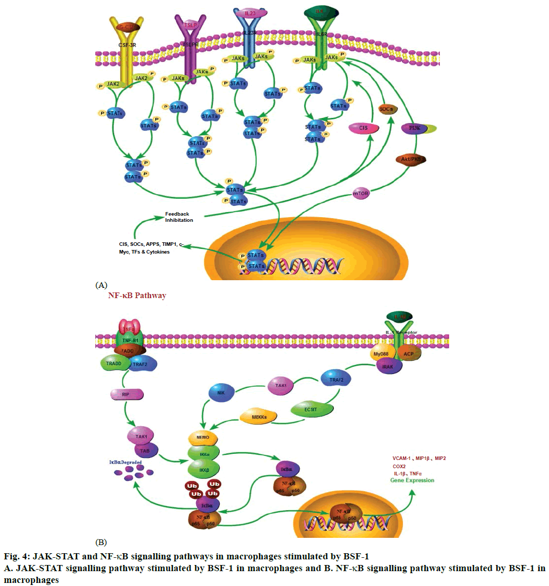
Through the KEGG pathway analysis it was realized that BSF-1 could induce several signalling pathways in macrophages to initiate an immune response, such as Janus kinase (JAK) and signal transducer and activator of transcription (STAT), NF-ҡB, TNF and MAPK signalling pathways. In this study, the data revealed that two major signalling pathways were induced, the JAKSTAT signalling pathway and the NF-ҡB signalling pathway (Figure 4).
Various ligands that could bind to cell surface receptors such as G-CSF (3766.5 fold), IL-6 (263.5 fold), IL-23 (79.8 fold), thymic stromal lymphopoietin (TSLP) (6.1 fold) all of which could activate associated JAKs and increase their kinase activity [16]. The tyrosine residues on the receptor were phosphorylated by activated JAKs, which could create SH2 domains in order to bind to proteins. STATs belonging to SH2 domains were gathered together by the receptor where their tyrosine residues were also phosphorylated by JAKs. STATs tyrosine residues could also be phosphorylated not only by the non-receptor tyrosine kinases in cytoplasm such as c-Src, but also by tyrosine kinases receptor such as the epidermal growth factor receptor [17]. Transcription of target genes was induced by these activated STATs, which constituted hetero- or homodimers and diverted to the cell nucleus [18].
Ligands such as TNFα (1127.9 fold) and IL-1β (7.3 fold) when bind to corresponding cell surface receptors can activate NF-ҡB signalling pathway. The products of such activation included TNFα (1127.9 fold), VCAM-1 (14.9 fold), IL-1β (7.3 fold). NF-ҡB was a transcription factor protein family containing five subunits which were NF-κB1, NF-κB2, RelA, RelB and c-Rel [19]. While in the resting state, NF-κB is combined with the inhibitory protein IκBα in cytoplasm. A great diversity of signals outside the cell bound to membrane receptors so as to activate the enzyme IκB kinase (IKK). The IκBα protein was phosphorylated by IKK, which led to the disintegration of the complex of NF-κB and IκBα. The free activated NF-κB was diverted into the nucleus to combine with specific sequences of DNA. The complex of NF-κB and DNA gathered other proteins such as coactivators and RNA polymerase to transcribe downstream DNA into mRNA, which was translated into protein resulting in a change of cell function [20].
Many researchers studied BSF-1 on human disease, but specific studies aimed at understanding the mechanism by which BSF-1 affected the immune system and the involved signalling pathways were seldom conducted. Therefore, the present investigation was conducted to find the signalling transduction pathways of BSF-1 activity on macrophages.
The DEAE-Sepharose fast flow column was used to extract and purify BSF-1, which was used to stimulate macrophages to get cytokines. Protein chip experiments were conducted to identify the secreted cytokines, which were 200 in number, including IL- 1β, IL-20, IL-23, TNF-α, and G-CSF. It turned out that there were 48 up-regulated cytokines, 73 downregulated cytokines and 79 unchanged cytokines. After analysis, it could be concluded that BSF-1 activated immune cells to secrete IL, which played a significant role in the maturation, activation, proliferation and immune regulation of the immune cells. To further study the mechanism of the BSF-1 stimulation of human immune system, all the factors in macrophages induced by BSF-1 were uploaded into the KEGG database in order to analyze the signalling pathway. By the KEGG analysis, it was found that BSF-1 could induce immune response of macrophages through a number of signalling pathways and there were two relatively important signalling pathways, the JAKSTAT and the NF-κB signalling pathways. These two signalling pathways are related to many diseases of the human body [21]. For instance, TNF-α, IL-6 and IL-1 are closely related to the pathogenesis of rheumatoid arthritis and joint destruction.
The JAK-STAT signalling pathway was one of the most important signalling transduction pathways found in recent years [22]. The JAK-STAT signalling cascade was made up of three main components, receptor, a JAK and two STAT proteins [23]. JAK is a family of nonreceptor tyrosine kinases, which transduced cytokinemediated signals via the JAK-STAT pathway. The STAT protein is a family of intracellular transcription factors. The JAK-STAT signalling pathway could transmit chemical signals from outside the cell to the nucleus leading to genes involved in immunity, proliferation [24], differentiation, apoptosis and oncogenesis transcribing and translating [25]. BSF-1 could promote macrophages secreting cytokines through JAK-STAT signalling pathway and had an effect on macrophage functions regulating the body's immune system, involved in cell proliferation, differentiation, survival, apoptosis, immune dysfunction and tumor formation.
These cytokines, G-CSF (3766.5 fold), IL-6 (263.5 fold), IL-23 (79.8 fold) and TSLP (6.1 fold), were found in the JAK-STAT signalling pathway in this study. G-CSF is a glycoprotein [26]. It is mainly produced by endothelial cells, macrophages, epidermal cells and fibroblasts stimulated by endotoxin, TNFα, IFN-γ and so on [27]. G-CSF mainly acted on the proliferation, differentiation and activation of neutrophil and hematopoietic cells. IL-6 is mainly secreted by activated T cells and fibroblasts, which can make B cell precursors, become cells that can produce antibodies. IL-6 combined with colony stimulating factor can promote the growth and differentiation of primordial bone marrow-derived cells and enhance the cleavage function of natural killer cells. IL-23 is produced by activated dendritic cells and macrophages, which is a cytokine, belonging to the IL-12 family. IL-23 can affect the differentiation of primitive Th cells into Th1 cells and regulate the production of Th1 cells. TSLP is a signalling molecule secreted by non-hematopoietic cells such as fibroblasts, epithelial cells and different types of stromal or stromal-like cells. It is a compound that has a capable of resulting in a strong immune response.
In addition, with the discovery of the connection between the NF-ҡB signalling pathway and some diseases such as cancer, asthma, muscular dystrophy, the NF-ҡB signalling pathway will be more concerned by people. Moreover, blocking the NF-ҡB signalling pathway to prevent and control some diseases will become another focus of future research. NF-ҡB is a protein complex, which controls the transcription of DNA, cytokine production and cell survival. The main function of NF-ҡB is the regulation of cell proliferation and apoptosis, immune and inflammatory reaction, which plays a very remarkable part in macrophages activation [28].
These cytokines, TNFα (1127.9 fold), VCAM-1 (14.9 fold), IL-1β (7.3 fold), were found in the NF- ҡB signalling pathway in this study. TNFα is secreted chiefly by activated macrophages. TNFα, of which the primary role is in the regulation of immune cells, is a cytokine capable of directly killing tumor cells without significant toxicity to normal cells. It is one of the most potent bioactivity factors to kill tumours directly. Vascular cell adhesion molecule 1 (VCAM-1) acts as a cell adhesion molecule produced by activated macrophages, fibroblasts, dendritic cells and so on. VCAM-1 not only regulated cadmium adhesion and vascular permeability [29], but also mediated neutrophil aggregation, infiltration to reach inflammatory site to clear pathogens [30]. IL-1β belongs to the IL-1 family of cytokines, which is produced by activated macrophages. IL-1β is an important mediator of the inflammatory response, and participates in a mass of cellular activities, including cell proliferation, differentiation, and apoptosis.
In brief, BSF-1 with the unique structure could activate immune cells to secrete cytokines, which can regulate the body's antitumor immunity and other related aspects, such as IL and tumor necrosis factor. In this study, the cytokines fulfilled a valuable function in two major signalling pathways, which were JAK-STAT signalling pathway and NF-ҡB signalling pathway through KEGG analysis. Through the study, it was found that the edible fungus polysaccharides could stimulate immune cells to secrete immune factors, which could provide a scientific basis for targeting sugar immunization preparations.
Acknowledgements
This project was supported by the Science and Technology Support Project of Sichuan Province (2018JY0087 and 2018NZ0055), the Cultivate Major Projects of Sichuan Province (16CZ0018), Nanchong science and Technology Bureau of Sichuan Province (16YFZJ0043), Talent Program of China West Normal University (17YC328,17YC136,17YC329), National Training Project of China West Normal University (17c039) and Innovative Team Project of China West Normal University (CXTD2017-3).
References
- Qin JZ, Chen M, Chen H, Lv JL. Prospect and current studies on edible and pharmaceutical fungi polysaccharides. Edible Fungi China 2004;23(2):6-9.
- O’Byrne KJ, Dalgleish AG. Chronic immune activation and inflammation as the cause of malignancy. Br J Cancer 2001;85(4):473-83.
- Le YY, Zhou Y, Iribarren P, Wang JM. Chemokines and chemokine receptors: their manifold roles in homeostasis and disease. Cell Mol Immunol 2004;1(2):95-104.
- Lieberman LA, Hunter CA. The role of cytokines and their signalling pathways in the regulation of immunity to Toxoplasma gondii. Int Rev Immunol 2002;21(4-5):373-403.
- Khazen W, M’bika JP, Tomkiewicz C, Benelli C, Chany C, Achour A, et al. Expression of macrophage-selective markers in human and rodent adipocytes. FEBS Lett 2005;579(25):5631-4.
- Hou YL, Ding X, Hou WR, Song B, Wang T, Wang F, et al. Pharmacological evaluation for anticancer and immune activities of a novel polysaccharide isolated from Boletus speciosus Frost. Mol Med Rep 2014;9(4):1337-44.
- Ding X, Feng S, Cao M, Li M, Tang J, Guo C, et al. Structure characterization of polysaccharide isolated from the fruiting bodies of Tricholoma matsutake. Carbohydr Polymers 2010;81(4):942-7.
- Ding X, Hou YL, Hou WR. Structure elucidation and antioxidant activity of a novel polysaccharide isolated from Boletus speciosus Forst. Int J Biol Macromol 2012;50(3):613-8.
- Hou Y, Liu L, Ding X, Zhao D, Hou W. Structure elucidation, proliferation effect on macrophage and its mechanism of a new heteropolysaccharide from Lactarius deliciosus Gray. Carbohydr Polym 2016;5(152):648-57.
- Zha X, Luo J, Jiang S. Induction of Immunomodulating Cytokines by Polysaccharides from Dendrobium huoshanense. Pharma Biol 2007;45(1):71-6.
- Zhang SM, Qiu J, Tian F, Guo XK, Zhang FQ, Huang QF. Corrosion behavior of pure titanium in the presence of Actinomyces naeslundii. J Mater Sci Mater Med 2013;24(5):1229-37.
- Brocker C, Thompson D, Matsumoto A, Nebert DW, Vasiliou V. Evolutionary divergence and functions of the human interleukin (IL) gene family. Human Genom 2010;5(1):30-55.
- Bakir-Gungor B, Sezerman OU. A new methodology to associate SNPs with human diseases according to their pathway related context. PLoS One 2011;6(10):e26277.
- Jin T, Li X, Zhang J, Wang H, Geng T, Li G, et al. Genetic association between selected cytokine genes and glioblastoma in the Han Chinese population. BMC Cancer 2013;13:236.
- Chen SC, Mehrad B, Deng JC, Vassileva G, Manfra DJ, Cook DK, et al. Impaired pulmonary host defense in mice lacking expression of the CXC chemokine lungkine. J Immunol 2001;165(5):3362-8.
- Brooks AJ, Dai W, O'Mara ML, Abankwa D, Chhabra Y, Pelekanos RA, et al. Mechanism of activation of protein kinase JAK2 by the growth hormone receptor. Science 2014;344:1249783.
- Levy DE, Darnell JE. Signalling: Stats: transcriptional control and biological impact. Nat Rev Mol Cell Biol 2002;3(9):651-62.
- Hebenstreit D, Horejs-Hoeck J, Duschl A. JAK/STAT-dependent gene regulation by cytokines. Drug News Perspect 2005;18(4):243-9.
- Nabel GJ, Verma IM. Proposed NF-kappa B/I kappa B family nomenclature. Genes Dev 1993;7(11):2063.
- Perkins ND. Integrating cell- signalling pathways with NF-kappaB and IKK function. Nat Rev Mol Cell Biol 2007;8(1):49-62.
- Harel S, Higgins CA, Cerise JE, Dai Z, Chen JC, Clynes R, et al. Pharmacologic inhibition of JAK-STAT signalling promotes hair growth. Sci Adv 2015;1(9):e1500973.
- Song X, Zhang Z, Wang S, Li H, Zuo H, Xu X, et al. A Janus Kinase in the JAK/STAT signalling pathway from Litopenaeus vannamei is involved in antiviral immune response. Fish Shellfish Immunol 2015;44(2):662-73.
- Aaronson DS, Horvath CM. A road map for those who don't know JAK-STAT. Science 2002;296(5573):1653-5.
- de Freitas RM, da Costa Maranduba CM. Myeloproliferative neoplasms and the JAK/STAT signalling pathway: an overview. Rev Bras Hematol Hemoter 2015;37(5):348-53.
- Duzagac F, Inan S, Ela Simsek F, Acikgoz E, Guven U, Khan SA, et al. JAK/STAT pathway interacts with intercellular cell adhesion molecule (ICAM) and vascular cell adhesion molecule (VCAM) while prostate cancer stem cells form tumor spheroids. J BUON 2015;20(5):1250-7.
- Ebihara Y, Xu MJ, Manabe A, Kikuchi A, Tanaka R, Nakahata T, et al. Exclusive expression of G-CSF receptor on myeloid progenitors in bone marrow CD34+ cells. Br J Haematol 2000;109(1):153-61.
- Sano E, Ohashi K, Sato K, Kashiwagi M, Joguchi A, Naruse N. A possible role of autogenous IFN-β for cytokine productions in human fibroblasts. J Cell Biochem 2007;100(6):1459-76.
- Kleniewska P, Piechota-Polanczyk A, Michalski L, Michalska M, Balcerczak E, Zebrowska M, et al. Influence of block of NF-kappa B signalling pathway on oxidative stress in the liver homogenates. Oxid Med Cell Longev 2013;2013:308358.
- Sarelius IH, Glading AJ. Control of vascular permeability by adhesion molecules. Tissue Barriers 2015;3(1-2):e985954.
- Qiu HN, Wong CK, Chu IM, Hu S, Lam CW. Muramyl dipeptide mediated activation of human bronchial epithelial cells interacting with basophils: a novel mechanism of airway inflammation. Clin Exp Immunol 2013;172(1):81-94.


