- *Corresponding Author:
- Zhenyu Xu
Department of Emergency Medicine, Seventh People's Hospital of Shanghai University of Traditional Chinese Medicine, Pudong, Shanghai 200137, China
E-mail: xzy2062@163.com
| This article was originally published in a special issue, “Recent Progression in Pharmacological and Health Sciences” |
| Indian J Pharm Sci 2024:86(2) Spl Issue “142-148” |
This is an open access article distributed under the terms of the Creative Commons Attribution-NonCommercial-ShareAlike 3.0 License, which allows others to remix, tweak, and build upon the work non-commercially, as long as the author is credited and the new creations are licensed under the identical terms
Abstract
Mitochondria play an important role in acute kidney injury. Yi Shen Pai Du formula is an effective traditional Chinese medicine formula for the clinical treatment of acute kidney injury. However, this study aimed to explore and investigate the mechanism of Yi Shen Pai Du formula on acute kidney injury. C57BL/6 mice were randomly divided into the control group, the acute kidney injury model group, and the Yi Shen Pai Du formula group (n=25). The levels of serum urea nitrogen, creatinine, and the expressions of reactive oxygen species, superoxide dismutase, and malondialdehyde in the mice were examined. The mitochondrial deoxyribonucleic acid copy number was detected by quantitative polymerase chain reaction. We report that the serum urea nitrogen and creatinine levels significantly increased in the Yi Shen Pai Du formula group. The reactive oxygen species expression was elevated while the expression of reactive oxygen species was decreased, and the expression of malondialdehyde was elevated. The oxidative stress biomarkers, such as malondialdehyde expression enhanced while superoxide dismutase expression was reduced. In addition, the mitochondrial deoxyribonucleic acid copy number level was significantly increased. Furthermore, the transmission electron microscopy showed that the number of mitochondria in the renal tubular epithelial cells of the mice of the Yi Shen Pai Du formula group was significantly increased compared with that in the model group, and the morphology of the mitochondrial cells was more intact. Our results led us to speculate that the Yi Shen Pai Du formula could effectively promote renal function recovery in mice with acute kidney injury and alleviate the degree of transformation from acute kidney injury to chr onic kidney disease.
Keywords
Yi Shen Pai Du formula, mitochondria, acute kidney injury, reactive oxygen species, integrative medicine
Acute Kidney Injury (AKI) is a common complication in clinical practice. It is a clinical syndrome characterized by acute tubular injury due to a sudden decline in renal function caused by pre-renal, renal or post-renal factors, including ischemia, infection or nephrotoxic drugs. Numerous studies have shown that repeated and severe AKI increases the risk of developing Chronic Kidney Disease (CKD) and ultimately irreversibly progresses to End-Stage Renal Disease (ESRD)[1-3]. Recently, several studies have concluded that during the development of AKI to CKD, mitochondrial metabolism of renal tubular epithelial cells is abnormal due to various blows. Damage to the mitochondria leads to the inhibition of aerobic glycolysis and the reduction of Adenosine Triphosphate (ATP) synthesis, which releases reactive oxygen radicals and other substances, resulting in necrosis or apoptosis of renal tubular epithelial cells and the loss of differentiation and regeneration ability[4,5]. Studies have shown that inhibiting mitochondrial membrane permeability transition pore opening can reduce renal injury in ischemia-reperfusion[6].
The protection, repair and regeneration of renal tubular mitochondria play a crucial role in inhibiting the transition from AKI to CKD, and the improvement of mitochondrial function may become an effective target for treating related diseases. The formula "benefiting kidney and Laxing turbid formula" is an empirical formula summarized by the Department of Nephrology, Yueyang Hospital of Integrative Medicine, and Shanghai University of Traditional Chinese Medicine, which has been used for a long time in clinical practice. In this study, it was experimentally proved that Yi Shen Pai Du formula can repair or regenerate renal tubular mitochondria in AKI, reduce the synthesis of extracellular matrix in renal fibroblasts, and thus inhibit the occurrence of renal fibrosis. This study aims to use Chinese medicine to reduce the incidence of AKI and delay its development into CKD, fully reflecting the advantages of Chinese medicine.
Materials and Methods
Animal study:
This experiment was approved by the Ethics Committee for Animal Experimentation of Shanghai University of Traditional Chinese Medicine. The experimental animals were 75 male C57BL/6 mice (8 w old) Specific Pathogen Free (SPF) grade, weighing about 20 g, purchased from Shanghai Sipul-Bikai Laboratory Animal Co., Ltd., Certificate of Conformity No: 20170005051199. In feeding conditions; room temperature maintained at 20°-24°, relative humidity was 45 %-50 %, artificial light from 7:00 AM-7:00 PM. During the experiment, 5 mice per cage could drink and eat freely, and the animal centre unified pure water and feed.
The mice were randomly divided into three groups; the control group, the AKI model group, and the Yi Shen Pai Du formula group, with 25 mice in each group. Each group was given drugs (frequency of administration, number of times/day) starting 7 d before modelling and ending when the Yi Shen Pai Du formula group was gavaged with Yi Shen Pai Du formula, and the control and model groups were replaced with the same amount of physiological saline.
AKI modelling:
According to the early reported procedure constructed the ischemia/reperfusion AKI model[7]; C57BL/6 mice were shaved and sterilized, anesthetized. The abdominal cavity was opened, and miniature arterial clips were applied to clip the bilateral renal arteries of the mice. The colour of the kidneys was observed to change to a purplish-black colour to indicate the success of ischemia. After 26- 30 min of clamping, the arterial clips were released, and the colour of the kidneys was observed to change rapidly to bright red, indicating that the model had been successfully constructed, and the openings were closed in two layers and sterilized. The vital signs of the mice were closely observed during the procedure, and the mice were returned to clean cages after awakening. The status of the mice was monitored periodically.
In the control group, the AKI mice model group, and the Yi Shen Pai Du formula group, the colour change of the kidneys was observed after modelling to determine the recovery of blood flow, and 5 mice were randomly taken at 5 min and 60 min for blood sampling and execution to obtain kidney tissues. The remaining mice in each group were collected in metabolic cages for 24 h at 24 h, 48 h, and 4 w, and 5 mice were randomly selected for blood collection, serum extraction, and execution to obtain kidney tissues.
Preparation of Yi Shen Pai Du formula:
Yi Shen Pai Du formula consists of raw Astragalus 30 g, cooked rhubarb 30 g, Tai Zi ginseng 15 g, raw rhubarb 30 g, Huang Jing 15 g, Xian Ling Zhen 30 g, safflower 3 g, leeches 3 g, granules formulated to be administered at a concentration of 150 mg/d, dissolved in saline. The granules were purchased from the Chinese herbal pharmacy of the Pharmacy Department of the Shanghai Seventh People's Hospital, the manufacturer, and each Chinese herbal granule batch number.
Biochemical analysis:
The blood serum samples were used to examine for creatinine and urea nitrogen using blood creatinine (C011-1, Nanjing Jianjian Institute of Bioengineering) and urea nitrogen (C013-2, Nanjing Jianjian) test kits. In addition, the levels of Reactive Oxygen Species (ROS), Superoxide Dismutase (SOD) and Malondialdehyde (MDA) in renal tissues were detected by referring to the instructions of ROS (E004, Nanjing Jianjian), total (SOD, A001- 1, Nanjing Jianjian) and (MDA, A003-1-2, Nanjing Jianjian) assay kits.
Reverse Transcription-quantitative Polymerase Chain Reaction (RT-qPCR) for mitochondrial Deoxyribonucleic Acid (DNA) copy number detection:
Kidney tissues were cut and fully ground in a mortar by adding liquid nitrogen. About 0.1 g of tissue powder was added to a 1.5 ml centrifuge tube, and tissue DNA was extracted using the tissue DNA extraction kit (DP304, Tiangen Biochemical Technology). The extracted DNA was amplified using the SYBR Green qPCR Master Mixes kit (Thermo Fisher) on a fluorescent qPCR instrument (ABI-7300). Samples Cycle threshold (Ct) value was defined as the number of cycles in which the fluorescence signal in the reaction tube reached a set threshold. Cyclooxygenase-1 (COX-1) is a mitochondrial gene; the 36B4 gene is a single copy number gene in vivo. Firstly, the COX-1 and 36B4 2-Ct values ratio was calculated (T/S), and then the T/S value of the experimental group was divided by the average T/S value of the control group to obtain the relative T/S value. When the relative T/S value=1, it means that the mitochondrial copy number of the experimental group is the same as that of the control group, relative T/S value <1 means that the mitochondrial copy number decreases, and relative T/S value >1 means that the mitochondrial copy number increases.
Transmission electron microscopy:
Kidney tissues were taken and fixed in 4 % glutaraldehyde solution for 24 h and then rinsed using Phosphate Buffer Solution (PBS) at pH 7.4, followed by 1 % osmium fixation for 1 h and then rinsed again. Alcohol gradient dehydration and embedding with epoxy resin 618 were used to make transmission electron microscopy specimens, which were sectioned and stained with uranium-lead double metal staining, and finally, the ultrastructure of the stage was observed under a transmission electron microscope (JEM-1230 transmission electron microscope, Japan); all electron microscopic photographs were recorded using a colour image recorder input device (JVC ky-F30B 3-CCD, Japan), and then the image analysis system (KONTRON image analysis system) was used for the analysis. An image analysis system (KONTRON IBAS 2.0 German) was used to analyse the pictures. Changes in mitochondrial morphology and distribution characteristics were also observed on a computer monitor.
Statistical analysis:
Statistical Package for the Social Sciences (SPSS) 21.0 statistical software was used for data statistics and analysis. Shapiro-Wilk normality test was used for the normality test, Levene’s test was used for checking the Chi-square (χ2) of variance, and mean±Standard Deviation (SD) was used for the statistical description of the measurement data conformed to normal distribution and χ2. For intra-group comparisons, Analysis of Variance (ANOVA) with repeated measures was used, and for inter-group comparisons, paired t-tests were used. The median was used to describe the data that did not conform to normal distribution, and the nonparametric rank sum test was used; frequency counts and constitutive ratios characterized the count data, and the χ2 test was used to analyse the indexes that were not in order; and the Kruskal-Wallis H test was used to compare the data between groups if the analyzed indexes were unidirectional ordered. All statistical tests were two- sided; the difference was considered statistically significant at p≤0.05.
Results and Discussion
The most often used clinical markers to assess renal function are serum urea nitrogen and creatinine. To investigate the effect of Yi Shen Pai Du formula in AKI mice, we evaluate the expression of serum urea nitrogen and creatinine levels at different time points. The results showed that the levels of serum urea nitrogen and creatinine were not remarkably changed in the AKI mice model and Yi Shen Pai Du formula group at a 5 min time point, whereas significantly reduced in the Yi Shen Pai Du formula group compared with the AKI mice model at 60 min, 24 h, 48 h, and 4 w time points (all, p<0.01) (Table 1 and Table 2). The result indicates that the Yi Shen Pai Du formula can reduce the degree of renal function damage in AKI and alleviate the degree of transformation from AKI to CKD.
| Time-point | n | Group | F | p | ||
|---|---|---|---|---|---|---|
| Control | AKI model | Yi Shen Pai Du formula | ||||
| 5 min | 5 | 6.50±1.15 | 6.50±1.60 | 6.98±1.52 | 0.181 | >0.05 |
| 60 min | 5 | 6.46±0.92 | 11.21±0.80 | 8.32±1.15 | 23.855 | <0.01 |
| 24 h | 4 | 8.27±0.86 | 14.10±1.96 | 9.56±1.34 | 19.36 | <0.01 |
| 48 h | 5 | 6.81±0.74 | 14.11±1.56 | 9.44±0.77 | 41.391 | <0.01 |
| 4 w | 5 | 6.15±1.14 | 14.00±2.20 | 9.20±0.52 | 31.617 | <0.01 |
| F | 0.181 | 19.186 | 4.62 | |||
| p | >0.05 | <0.01 | <0.05 | |||
Table 1: Comparison of Serum Urea Nitrogen Levels at Different Time Point in Different Mice Model (μmol/l)
| Time-point | n | Group | F | p | ||
|---|---|---|---|---|---|---|
| Control | AKI model | Yi Shen Pai Du formula | ||||
| 5 min | 4 | 27.78±4.24 | 27.69±7.58 | 31.46±6.34 | 0.94 | >0.05 |
| 60 min | 5 | 26.18±5.67 | 80.05±10.12 | 53.49±4.77 | 48.53 | <0.01 |
| 24 h | 5 | 31.64±12.47 | 120.73±14.45 | 70.26±13.72 | 42.69 | <0.01 |
| 48 h | 5 | 40.50±8.96 | 97.57±14.97 | 75.15±12.05 | 19.67 | <0.01 |
| 4 w | 5 | 35.41±8.15 | 102.84±15.78 | 60.65±6.13 | 35.37 | <0.01 |
| F | 0.89 | 37.58 | 16.94 | |||
| p | >0.05 | <0.01 | <0.05 | |||
Table 2: Comparison of Serum Creatinine Levels at Different Time Points in Different Mice Models (μmol/l)
ROS, MDA and SOD are considered important pathophysiologic factors in the progression of renal disease. We did not observe a significant change in the AKI mice model and Yi Shen Pai Du formula group in the levels of ROS and MDA at 5 min time points and SOD at 5 min and 60 min time points. On the other hand, ROS and MDA levels were potentially decreased at 60 min, 24 h, 48 h, and 4 w time points, whereas SOD levels increased at 24 h, 48 h, and 4 w time points in the Yi Shen Pai Du formula group compared with the AKI mice model (all, p<0.01) (Table 3-Table 5). The results demonstrate that the Yi Shen Pai Du Formula considerably reduced ROS and MDA levels, and the level of SOD increased in AKI mice.
| Time-point | n | Group | F | p | ||
|---|---|---|---|---|---|---|
| Control | AKI model | Yi Shen Pai Du formula | ||||
| 5 min | 5 | 1062.38±72.05 | 989.66±56.31 | 1040.64±20.28 | 2.38 | >0.05 |
| 60 min | 5 | 951.80±90.68 | 2304.10±81.07 | 1447.43±99.74 | 283.7 | <0.01 |
| 24 h | 5 | 1001.24±107.87 | 1953.29±284.62 | 1322.33±180.03 | 28.14 | <0.01 |
| 48 h | 5 | 1017.35±87.44 | 2031.11±119.66 | 1342.77±140.35 | 96.45 | <0.01 |
| 4 w | 5 | 935.46±161.25 | 2054.52±340.46 | 1176.38±124.52 | 33.06 | <0.01 |
| F | 1.11 | 29.14 | 8.08 | |||
| p | >0.05 | <0.01 | <0.01 | |||
Table 3: Comparison of Renal Tissue ROS Levels at Different Time Points in Different Mice Models (RLU/mgprot)
| Time-point | n | Group | F | p | ||
|---|---|---|---|---|---|---|
| Control | AKI model | Yi Shen Pai Du formula | ||||
| 5 min | 5 | 12.37±0.94 | 12.03±1.42 | 12.39±1.11 | 0.15 | >0.05 |
| 60 min | 5 | 10.39±1.26 | 21.52±1.72 | 13.99±3.27 | 31.96 | <0.01 |
| 24 h | 5 | 12.22±2.57 | 23.95±3.29 | 12.97±3.29 | 22.87 | <0.01 |
| 48 h | 5 | 12.36±1.95 | 22.91±0.91 | 15.27±1.60 | 66.35 | <0.01 |
| 4 w | 5 | 11.11±2.53 | 24.21±4.61 | 11.02±2.02 | 27.24 | <0.01 |
| F | 1.04 | 17.27 | 2.196 | |||
| p | >0.05 | <0.01 | >0.05 | |||
Table 4: Comparison of Renal Tissue MDA Levels at Different Time Points in Different Mice Models
| Time-point | n | Group | F | p | ||
|---|---|---|---|---|---|---|
| Control | AKI model | Yi Shen Pai Du formula | ||||
| 5 min | 5 | 60.07±4.17 | 52.25±4.07 | 51.00±4.18 | 7.05 | >0.05 |
| 60 min | 5 | 58.54±7.30 | 17.92±4.75 | 18.01±4.59 | 119.7 | >0.05 |
| 24 h | 5 | 56.96±5.74 | 10.31±2.91 | 31.06±8.00 | 77.76 | <0.01 |
| 48 h | 5 | 64.98±5.64 | 13.09±6.96 | 35.58±7.26 | 76.36 | <0.01 |
| 4 w | 5 | 56.13±10.12 | 16.67±7.70 | 38.53±12.71 | 75.25 | <0.01 |
| F | 1.474 | 47.32 | 11.3 | |||
| p | >0.05 | <0.01 | <0.01 | |||
Table 5: Comparison of Renal Tissue SOD Levels (U/mgprot) at Different Time Points in Different Mice Models
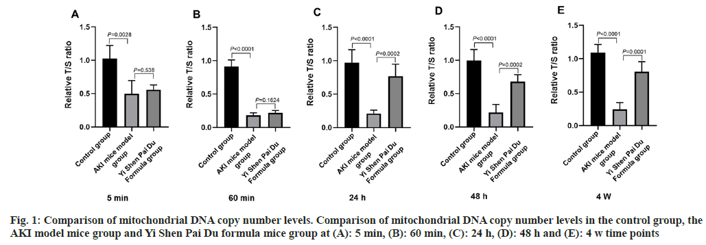
We performed RT-qPCR analysis to investigate the effect of Yi Shen Pai Du formula on mitochondrial DNA copy number levels. The results showed that the mitochondrial DNA copy numbers were significantly reduced in the AKI mice model compared with the control group at 5 min, 60 min, 24 h, 48 h, and 4 w time points. On the other hand, except at 5 min and 60 min time points, the mitochondrial DNA copy numbers were dramatically increased in the Yi Shen Pai Du formula group at 24 h, 48 h, and 4 w time points compared with the AKI mice model (fig. 1A-fig. 1E). The results led us to speculate that the Yi Shen Pai Du formula can significantly reduce mitochondria damage in AKI mice and potentially restore the number of mitochondria with time.
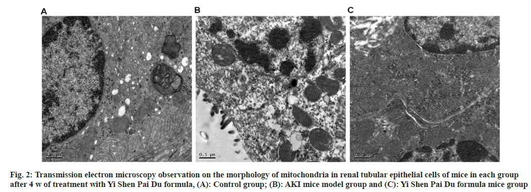
After 4 w of treatment with Yi Shen Pai Du formula, transmission electron microscopy was used to observe the morphological changes of mitochondria of renal tubular epithelial cells, and it was found that mitochondria of renal tubular epithelial cells of rats in the control group were abundant and normal in morphology. The cytoplasm was dense, and the mitochondrial cristae were intact (fig. 2A). In the AKI mice model group, the mitochondria of renal tubular epithelial cells and cytoplasm were lost, the mitochondrial cristae were broken, and many vacuoles appeared in the cytoplasm (fig. 2B). The number of mitochondria in the renal tubular epithelial cells of mice in the group of Yi Shen Pai Du formula was slightly less than that in the control group, the morphology was normal, with part of the cytoplasm being less dense, and part of the mitochondrial cristae being fractured (fig. 2C). Our results demonstrated that the Yi Shen Pai Du formula could significantly reduce the AKI damage in mitochondria and maintain the number and morphology of mitochondria.
In this experiment, we investigated the protective effect of Yi Shen Pai Du formula on acute renal ischemia-reperfusion injury. The results showed that Yi Shen Pai Du formula significantly improved the renal function of the AKI model mice, reduced the structural damage of renal tubules, and decreased the phenomena of degeneration, necrosis and inflammatory cell infiltration in the drug intervention group. Compared with the AKI model mice, the serum MDA level decreased and increased the SOD level in the Yi Shen Pai Du formula mice group, suggesting that the protective mechanism of Yi Shen Pai Du formula against AKI is to regulate the production and release of ROS through the changes in mitochondrial dynamics, thus reducing the oxidative stress injury.
AKI is one of the most common complications in severely life-threatening emergency medicine admissions. Inadequate renal blood supply is the leading clinical cause of AKI[8,9]. Renal ischemia leads to renal microvascular changes and mitochondrial metabolic disturbances, increasing ROS[10,11]. Reperfusion induces excessive ROS production, and the accumulation of ROS further aggravates renal injury and leads to oxidative stress[12,13]. After oxidative stress induced by ischemia-reperfusion, the kidney protects itself through redox-sensitive mitochondrial enzymatic reactions[14,15].
Mitochondria are the power factories of the cell. Based on energy requirements, neural stem cell mitochondria regulate number, intracellular localization, energy metabolism, production of ROS, and removal of damaged mitochondria through mitochondrial biogenesis, division, and fusion, which changes shape, number, and complexation[16]. Studies have shown that renal tubular cells are rich in mitochondria and that tubular reabsorption and secretory functions depend on mitochondrial oxidative phosphorylation for energy[17,18]. Mitochondria play an essential role in AKI. In AKI, there is mitochondrial swelling, impaired dynamic homeostasis, and changes in mitochondrial membrane permeability, deficient mitochondrial production, decreased productivity and increased ROS production. Similarly, measures such as regulating mitochondrial morphology, controlling the opening of the mitochondrial membrane permeability transition pore, promoting mitochondrial production and using mitochondria-targeted antioxidants can effectively protect the integrity of mitochondrial morphology and function, thereby protecting renal function[19,20].
CKD does not have a name in Chinese medicine. According to its clinical symptoms, signs can be divided into edema, deficiency labor, kidney wind, Guan Ge and other diseases. Its pathogenesis is blood stagnation and kidney deficiency. The main ingredients of Yi Shen Pai Du formula are rhubarb, Tai Zi ginseng, Astragalus, Huang Jing, Xian Ling spleen, saffron and leech. Among them, rhubarb cleanses the evils of filth in the stagnant blood, Tai Zi ginseng and Astragalus replenish the vital energy and benefit the essential energy and blood, Huang Jing nourishes the kidney, Xian Ling Zhen spleen warms up the necessary energy, and saffron and leech break up the blood and remove the blood stasis, which benefits the kidneys and eliminates the turbid effect. The above drugs complement each other; Gu Yuan eliminates blood stasis, activates meridians and collaterals, and works together to reduce renal ischemia-reperfusion injury. It has been experimentally confirmed that rhubarb and Astragalus has antioxidant, anti-inflammatory and lipid metabolism multiple pharmacological effects[21-23].
In conclusion, Yi Shen Pai Du formula has a specific protective effect on the renal function of ischemia- reperfusion induced AKI mice. It could alleviate the transformation from AKI to CKD.
Acknowledgements:
Jiaying Pan conceived and designed the experiments, performed the experiments, analyzed and interpreted the data, and wrote the paper. Jinmiao Ma, Yubao Chen, and Ziqing Zhang analyzed and interpreted the data. Zhenyu Xu conceived and designed the experiments and contributed reagents, supervised materials, analysis tools or data.
Funding:
This study was supported by Shanghai Municipal Health Commission Scientific Research Project (No: 202040143); Pudong New Area Key Specialty Construction Project of Pudong Health and Family Planning Commission of Shanghai (Grant No: PWZzk2022-15) and Shanghai Municipal Seventh People’s Hospital “Beidou Star” Talent Training Plan Project (No: BDX2021-03).
Conflict of interests:
The authors declared no conflict of interest.
References
- Chawla LS, Amdur RL, Amodeo S, Kimmel PL, Palant CE. The severity of acute kidney injury predicts progression to chronic kidney disease. Kidney Int 2011;79(12):1361-9.
[Crossref] [Google Scholar] [PubMed]
- Coca SG, Singanamala S, Parikh CR. Chronic kidney disease after acute kidney injury: A systematic review and meta-analysis. Kidney Inter 2012;81(5):442-8.
[Crossref] [Google Scholar] [PubMed]
- Wald R, Quinn RR, Adhikari NK, Burns KE, Friedrich JO, Garg AX, et al. Risk of chronic dialysis and death following acute kidney injury. Am J Med 2012;125(6):585-93.
[Crossref] [Google Scholar] [PubMed]
- Szeto HH, Liu S, Soong Y, Seshan SV, Cohen-Gould L, Manichev V, et al. Mitochondria protection after acute ischemia prevents prolonged upregulation of IL-1β and IL-18 and arrests CKD. J Am Soc Nephrol 2017;28(5):1437.
[Crossref] [Google Scholar] [PubMed]
- Chevalier RL. The proximal tubule is the primary target of injury and progression of kidney disease: Role of the glomerulotubular junction. Am J Physiol Renal Physiol 2016;311(1):F145-61.
[Crossref] [Google Scholar] [PubMed]
- Bopassa JC, Eghbali M, Toro L, Stefani E. A novel estrogen receptor GPER inhibits mitochondria permeability transition pore opening and protects the heart against ischemia-reperfusion injury. Am J Physiol Heart Circ Physiol 2010;298(1):H16-23.
[Crossref] [Google Scholar] [PubMed]
- Kramann R, Fleig SV, Schneider RK, Fabian SL, diRocco DP, Maarouf O, et al. Pharmacological GLI2 inhibition prevents myofibroblast cell-cycle progression and reduces kidney fibrosis. J Clin Invest 2015;125(8):2935-51.
[Crossref] [Google Scholar] [PubMed]
- Scholz H, Boivin FJ, Schmidt-Ott KM, Bachmann S, Eckardt KU, Scholl UI, et al. Kidney physiology and susceptibility to acute kidney injury: Implications for renoprotection. Nat Rev Nephrol 2021;17(5):335-49.
- Jun Wu. A preliminary study on the forms of cell death in acute kidney injury due to ischemia-reperfusion. Yujiang Med Coll 2022.
- Tarasenko AI, Alekseev AV, Maksimova SY. Mechanisms of kidney damage. Urologiia 2019;2:103-7.
- Qingying NI, Zhenyang HE, Yaxuan F, Ke Z, Bo Y. Overview of research on Chinese medicine for acute kidney injury. J Tradit Chin Med 2022;50(10):93-6.
- Su L, Zhang J, Gomez H, Kellum JA, Peng Z. Mitochondria ROS and mitophagy in acute kidney injury. Autophagy 2023;19(2):401-14.
[Crossref] [Google Scholar] [PubMed]
- Xia T, Zhang X, Li D, Liu W, Qin Z. Research progress of oxidative stress and NOD-like/Toll-like receptors in acute kidney injury. J Hainan Med Coll 2020;26(9):717-20.
- Zhang AH. Mitochondria: A new target for acute kidney injury treatment. Chin J Pharmacol Toxicol 2019;33(10):825.
- Zhang X, Agborbesong E, Li X. The role of mitochondria in acute kidney injury and chronic kidney disease and its therapeutic potential. Int J Mol Sci 2021;22(20):11253.
[Crossref] [Google Scholar] [PubMed]
- Tang C, Cai J, Yin XM, Weinberg JM, Venkatachalam MA, Dong Z. Mitochondrial quality control in kidney injury and repair. Nat Rev Nephrol 2021;17(5):299-318.
[Crossref] [Google Scholar] [PubMed]
- Jin H, Zhang Y, Ding Q, Wang SS, Rastogi P, Dai DF, et al. Epithelial innate immunity mediates tubular cell senescence after kidney injury. JCI Insight 2019;4(2):e125490.
[Crossref] [Google Scholar] [PubMed]
- Chen X, Ma Y, Nie J, Zhu F. Research progress on the repair mechanism of renal tubular epithelial cell damage in the transition from acute kidney injury to chronic kidney disease. J Capital Med Univ 2022;43(5):720-7.
- Pabla N, Bajwa A. Role of mitochondrial therapy for ischemic-reperfusion injury and acute kidney injury. Nephron 2022;146(3):253-8.
[Crossref] [Google Scholar] [PubMed]
- Hu M, Li X, Gao J. Mitochondrial dynamics-related proteins and myocardial ischemia-reperfusion injury and the progress of Chinese medicine intervention. Chin Patent Med 2023;3(23):1-6.
- Jia H, Lu C, Yuan B, Liu H, Wang T. Protective effects of rhubarb extract on acute kidney injury and effects on Keap1/Nrf2 signaling pathway in septic rats. J Pharm Anal 2021;41(6):979-85.
- Lei X, Wei H, Tan R, Wang L. Effects of Astragalus Panax ginseng combination on cisplatin-induced acute kidney injury in mice. Chin Patent Med 2022;44(4):1107-13.
- Huang P, Luo L, Su P. Research progress of Astragalus combined with rhubarb in the treatment of chronic renal failure. J Pract Clin Med 2018;22(17):126-8.


