- *Corresponding Author:
- Xiuhua Mao
Department of General Surgery, West China Health Care Hospital of Sichuan University, Chengdu 610041, China
E-mail: 53220009@qq.com
| Date of Received | 28 November 2020 |
| Date of Revision | 15 January 2021 |
| Date of Acceptance | 10 April 2021 |
| Indian J Pharm Sci 2021;83(2):362-370 |
This is an open access article distributed under the terms of the Creative Commons Attribution-NonCommercial-ShareAlike 3.0 License, which allows others to remix, tweak, and build upon the work non-commercially, as long as the author is credited and the new creations are licensed under the identical terms
Abstract
In China, the incidence rate and mortality rate of gastric cancer are in the second place among all tumors and the pathological type is mainly adenocarcinoma. The higher prevalence rate requires us to do more research. It is of great significance to study the antitumor effect of hydroxysafflor yellow pigment A in gastric adenocarcinoma. The aim of this study was to investigate the inhibitory effect of hydroxysafflor yellow pigment A on human gastric adenocarcinoma BGC-823 xenograft in nude mice. The animal model of human gastric adenocarcinoma BGC-823 was established in nude mice to carry out the experiment and then the transmission electron microscope was used to observe and analyze the experimental data. The results showed that on the 4th d after inoculation, there were more subcutaneous tumor masses in nude mice and on the 7th d, all the nude mice formed tumor blocks with the size of 0.4 cm to 0.8 cm and the tumorigenesis rate was 100 %. With the continuous administration, the tumor weight of the small dose group was only 0.88 g, which was much smaller than that of the model group. The average percentage of early apoptosis in low dose group was 2.48 % and that in model group was 0.44 %. In the low dose group, the volume of tumor cells became smaller and apoptotic bodies appeared. Therefore, it is of great significance to study the inhibitory effect of hydroxysafflor yellow pigment A on human gastric adenocarcinoma BGC- 823 xenograft in nude mice.
- *Corresponding Author:
- Xiuhua Mao
Department of General Surgery, West China Health Care Hospital of Sichuan University, Chengdu 610041, China
E-mail: 53220009@qq.com
| Date of Received | 28 November 2020 |
| Date of Revision | 15 January 2021 |
| Date of Acceptance | 10 April 2021 |
| Indian J Pharm Sci 2021;83(2):362-370 |
This is an open access article distributed under the terms of the Creative Commons Attribution-NonCommercial-ShareAlike 3.0 License, which allows others to remix, tweak, and build upon the work non-commercially, as long as the author is credited and the new creations are licensed under the identical terms
Keywords
Hydroxysafflor yellow pigment A, nude mice, gastric adenocarcinoma, BGC-823 transplanted tumor
Gastric cancer is the most common form of gastrointestinal cancer, their specific tumor pathogenesis is not entirely clear. About 95 % of gastric adenocarcinoma is differentiated according to the severity of histological differentiation and can be divided into well differentiated benign and poorly differentiated adenocarcinoma. Early diagnosis and early radical resection have important clinical significance for the prognosis of gastric cancer patients. Most chronic cases of early gastric cancer have occurred in the hospital, diagnosed late, early gastric cancer treatment method is limited and the prognosis is very bad. So, to explore the molecular biology of gastric cancer, the discovery of new molecular tumor markers clear all kinds of stomach cancer pathogenesis of gastric cancer and develop effective prevention strategies and effective treatment of gastric cancer is imminent. It is worth mentioning that the early symptoms of gastric cancer patients are atypical, the clinical manifestations are relatively hidden and the early diagnosis is difficult. Most patients are already in the middle and late stages of the disease when they are diagnosed and cannot be surgically removed.
Regulation of cell signal transduction of human cells for proliferation, apoptosis and cell lifecycle activities coordinated balance between internal and external environment and human cells, an abnormal signal transduction can lose the normal regulatory cell function, leading to cell proliferation imbalance and apoptosis, such that cells uncontrolled growth of malignant tumor occurrence and development[1]. At present, for patients with advanced disease, the comprehensive treatment and surgery methods for gastric cancer can mainly include surgical gastrointestinal resection, new adjuvant surgery, chemotherapy and re-adjuvant surgery, radiotherapy and chemotherapy, but it has repeatedly invaded using gastric laparoscopy and other organs has multiple intestinal lymph node metastases of gastric cancer patients. Surgery is often difficult to further embodiments gastrointestinal radical resection[2]. Selecting an effective treatment regimen depends on comprehensive evaluation before surgery, including lesion size, range, depth and the situation involving the stomach wall lymph nodes and distant metastasis, chronic and cause, clinical physiology of gastric lesion type, severity and differentiation of biological behavior of learning diagnosis is closely related[3].
Wang studied whether hydroxysafflor yellow pigment A (HSYA) could improve the dyskinesia and fluctuation induced by levodopa in rats. Levodopa (8 mg/kg) and Benderizines (15 mg/kg) were injected intraperitoneally. HSYA was injected intraperitoneally at the dose of 10 mg/kg[4]. Ikeguchi study found that the average apoptotic index (AI) of 97 tumors was 2.05 % (range: 0-11.31 %)[5]. Azuma analysis showed that 121 cases of Helicobacter pylori were positive and 46 cases were negative. In the control group, 36 cases were superficial gastritis and 85 cases were atrophic gastritis[6]. Ajani enrolled 49 patients and 43 patients can be evaluated. The pathological complete response rate was 26 %. At 1 y, the survival rate of patients with complete pathological response (82 %) was higher than that of patients with complete pathological response[7].
Compared with the previous HSYA on nude mice with human gastric adenocarcinoma xenograft BGC- 823 (human gastric cancer cells) research literature, innovative content herein is broadly divided into the following points: the first point is successfully established using five groups of human gastric adenocarcinoma BGC-823 nude mice xenograft model, tumor volume and mass detection dissected, analyzed and compared. The second point is to analyze the pathogenesis of tumor tissue through optical microscope and transmission electron microscope and compare the aggregation and arrangement of tumor cells in nude mice, chromatin and nucleolus. The third point is to detect the apoptosis and cell cycle of tumor cells in nude mice by flow cytometry. So, more scientific research was done to study the inhibitory effect of HSYA on human gastric adenocarcinoma BGC-823 xenograft in nude mice, which has great reference value.
At present, the world’s new technologies for the treatment of malignant tumors have been focused on in depth study of how to effectively block the angiogenesis in tumors. According to reliable reports, angiogenesis in tumors can supply a variety of tumor metabolism at the same time. How to effectively control the growth and proliferation of tumor cells in vascular mucosal endothelial cells has become a key factor, control point for tumor inhibition and tumor angiogenesis. The results showed that HSYA has strong anti-acute platelet, anti-acute myocardial ischemia, anti-thrombin induced inhibition of platelet aggregation, anti-inflammatory, anti-tumor and other activities. Therefore, it is a natural Chinese herbal medicine monomer with great research value. Some experimental studies have shown that HSYA can antagonize and inhibit cell proliferation and contraction during the development of capillary wall and smooth muscle epithelial cells[8].
HSYA may restrain degradation of von Hippel-Lindau (VHL) and p53 cell cytokine induction, so that low oxygen, effectively increase the proliferation rate of human hypoxia[9]. In addition, safflower medicine may be used for anti-cancer therapy. Another important reason is, there are many studies, in which it said the use of safflower medicine can effectively reduce it long term. In some, high altitude hypoxia conditions remain large and active malignant cells such as tumor cells may also have resistance to chemotherapy and it will cause a major source of early tumor recurrence and it can increase the uniform fluidity of local blood in patients with early malignant tumors. The chemical component in safflower that can restrain and promote the curative effect of anti-cancer drugs is mainly a dimethyl alcohol named sterols and it is not good for the inflammation, cancer and tumor lesions on the skin of the experimental nude mice. It has obvious tumor inhibition effect. In addition, 70 % of the safflower extracts cholesterol cervical cancer cells in vivo in mice, a mouse lymphosarcoma tumor cell lines of mouse lymphoma cell line and no significant effect inhibit the reaction, while in vitro experiments in mice, it also has no obvious inhibitory effect on leukemia cells. The application of safflower injection can significantly prevent and reduce the incidence of ischemic stroke. Its own special antioxidant activity also makes it effective to improve the evaluation of the electrophysiological function of neuronal limbs and increase the index of the electrophysiological function of the spinal cord. That is, it can effectively inhibit and regulate B-cell lymphoma 2 (Bcl-2) synthesis and expression level of the protein which is effective to inhibit apoptosis of epithelial cells in spinal cord neurons where neurons increased antioxidant effect.
Safflower injection preparation in the interior of the abdomen of nude mice experiment can be effectively reduced by the ligation of arterial thrombus formation which caused edema effect. Superoxide and nitric oxide may form proximities. Using this antioxidant method of inducing the large amounts of glutamic acid, which can greatly reduce neuronal apoptosis, precisely because HSYA has an important effect on the human body to inhibit neuronal apoptosis[10]. A further HSYA mitochondrion for ischemic heart disease and brain injuries also showed significant inhibitory effect mobility and protection. These indicate mitochondrial membrane fluidity therefore reduced degree of inhibition, thereby increasing mitochondrial respiratory function. Clinical observation Clinical observations have shown that the receptor for the stimulant HSYA is an important physiological mechanism for neuroepithelial cells to produce inhibitory effects, according to the researchers. HSYA Bcl-2 receptor may be adjusted to suppress the N-methyl-D-aspartate (NMDA) receptor body, plays an important role in nerve cells. According to reports, the administration of the stimulant HSYA in the body can significantly reduce the neuropathy of neuronal epithelial cells.
HSYA may have two way functions on the production of N-methyl-D-aspartate receptor 1 (NMDAR1) protein after cerebral ischemia, which may be due to the inhibition of NMDAR1 protein expression after cerebral ischemia. Because safflower yellow can reduce the apoptosis rate of brain cells, increase the expression of Bcl-2 gene, so as to reduce the apoptosis of nerve cells. Safflower yellow can also significantly improve the chemical function defects of brain cell behavior system and the edema of brain cells in rats. Similarly, another recent study showed that the extract of safflower has a certain dose effect relationship on scavenging superoxide, hydroxyl and 2,2-diphenyl-1- picrylhydrazyl (DPPH) in cells. In addition, the petal extract of cantharus tinctures was found to be effective in preventing cell dysfunction and cell damage induced by hydrogen peroxide reaction. HSYA is also considered to significantly reduce the collagen content of alkaline cells and increase the receptor cell activation of nucleic acid growth factor ligand and the production of antioxidants. Therefore, it is suggested that HSYA has a protective effect on inducing cytotoxicity.
The main function of HSYA is that it can effectively reduce the oxidative stress reaction of other cells and time of apoptosis, so as to prevent or reduce the necrosis of nerve cells caused by ischemia or reperfusion in other nerve cells, bone marrow and thus reduce the protein content of malondialdehyde (MDA). Another type of yellow amino acid A, which contains a hydroxyl group called carthamin, is a chalcone like organic compound because its organic chemical molecular structure can reduce oxidative stress. It contains multiple hydroxyl groups. It suggests that the antioxidant effect and pharmacological activity of flavin a, which is called safflower, is related to the interaction of these substances, because it is caused by free radical reaction antioxidant effect which is a kind of pharmacological activity factor that may cause many chronic blood diseases. Therefore, the effective pharmacological components of safflower yellow have strong antioxidant activity in human body. Therefore, we can selectively reduce the possible oxidative disorder caused by improving blood circulation by effectively inhibiting the occurrence of free radical antioxidant reaction. HSYA has been proved to have therapeutic effect on ischemic brain injury in rats. It has also shown obvious effect in improving the clinical score of functional brain defect of central nervous system and reducing the risk of acute brain injury in high incidence area of ischemic acute cerebral infarction in rats, which may be related to the inhibition of thrombosis and platelet aggregation. It has a beneficial effect on the regulation of hemorheology.
HSYA can remove hydroxyl group in blood and inhibit fenton damage to oxidase, which fully proves that it is an effective antioxidant active part of hydroxy saffron yellow[11]. Compared with other substances, HSYA and anhydrosafflor yellow B (SYB) has weak scavenging ability to hydroxyl radicals. The other antioxidant component of saffron yellow has strong antioxidant ability to directly inhibit and scavenge hydroxyl radicals. At the same time, because of saffron yellow containing two kinds of antioxidant mechanism, the use of saffron yellow as a whole is reasonable. In addition, HSYA, as one of the effective antioxidant substances in safflower, has potential development prospects.
A HSYA discovered by scientists may attenuate lymphoid encephalopathy induced brain injury in rats and selective change related functions, which may be related to regulation of nitric oxide pathway. Safflower injection as a pharmaceutical formulation is generally widely used in clinical. It has only been clinically proven which has effective protection and inhibit acute myocardial ischemia caused by acute adrenergics[12]. This is likely to be related to the reduction in the quality of the entire heart valve protective protein caused by the inflammatory toxic response of tumor necrosis factor alpha (TNF-α) and the reduction of its protein level and the increase of the Bcl-2 protein level in the heart tissue, make it effective. In the anti-myocardial ischemia experiment, rats with myocardial ischemia and the rabbits were injected with intravenous injection of safflower injection. The researchers then detected using epicardial electrocardiogram imaging techniques, detection of the effect on myocardial ischemia of safflower injection and relief and suppression effect. The effect of HSYA can reduce the resistance of the coronary artery and myocardium of experimental dogs and at the same time increase the nutritional blood flow of the coronary arteries and myocardium of experimental dogs. In the in vitro experiment, it has a mildly exciting effect on the heart blood vessels of toads and rabbits. HSYA can also effectively make the coronary and heart blood vessels of experimental dogs contract and dilate. Another study found that, safflower yellow can reduce myocardial injury in rats, the mechanism of which HSYA can reduce the lactate dehydrogenase (LDH) and ventricular tissue adenosine triphosphate (ATP) content, but also HSYA may improve myocardial swelling of mitochondria, to enhance membrane fluidity.
Gastric cancer is a common gastrointestinal malignant tumor in the world and in the region. Although the average incidence rate and mortality rate of gastric cancer in China is significantly lower than that in other regions, the average incidence rate and mortality rate of gastric cancer in China are relatively high[13]. The most common histological type of gastric cancer is adenocarcinoma. Surgical resection is an effective treatment to cure gastric cancer. Although great progress has been made in the treatment of gastric cancer in recent years, the survival time of patients is still not ideal because most patients are in the middle and late stage when they are found. Recurrence and metastasis of gastric cancer are the main causes of death in patients with gastric cancer. The biological characteristics of malignant tumor include uncontrolled cell growth, abnormal differentiation, invasion and metastasis. Invasion and metastasis are the most important characteristics of malignant tumor and the occurrence of invasion and metastasis is also the main cause of death of most patients. In fact, the process of tumor cell differentiation and metastasis is a gradual and diversified stage of cell growth and development. The invasion of malignant tumor cells in a primary tumor can make the cells gradually penetrate through the basement membrane and infiltrate into the surrounding local tissues to make the cells metastasize and grow, invade and reach the surrounding tissues, local blood vessels and lymph nodes with the blood and the tubal lumen continued to proliferate and infiltrate. The mechanism of tumor invasion and metastasis is a complex pathological process, which is regulated by many factors, including the change of gene expression which is related to tumor invasion, the process of outer surface differentiation of cell membrane, including the changes of cell structure of benign cell adhesion molecules, the degradation of extracellular matrix, the formation of vascular stromal cells and the epithelial mesenchymal transition (EMT) of malignant cells, the binding process of protein transformation in cytoplasmic cells.
In the research of traditional Chinese medicine, it is found that the prescription composed of angelica daturic, radix astragali, dandelion, scorpion, centipede and so on can inhibit the lymph node metastasis of gastric cancer by inhibiting the expression of Bcl-2 protein. Bazhen granule adjuvant chemotherapy can inhibit the lymphatic metastasis of gastric cancer by down regulating human tumor suppressor gene p53. In addition, it can regulate the expression of bispecific protein phosphatase; extracellular regulation of protein kinase pathway can effectively inhibit the proliferation, invasion and metastasis of gastric cancer cells and reduce the lymph node metastasis rate of gastric cancer. At present, there are reports of prescriptions consisting of red vine, Scutellaria, rhubarb, Scutellaria slices, woody, tangerine peel, dang shen slices, etc. and prescriptions consisting of dang shen slices, astragalus, Polygonatum, Atractylodes, yam, cocos, raw coix seed, tangerine peel, etc. used to treat patients with gastric cancer can alleviate the side effects of radiotherapy and chemotherapy and significantly enhance the immune function of patients and improve their quality of life. Through clinical observation, it is verified that the prescriptions consisting of Codonopsis, Fried Atractylodes, Psoralen, Cuscuta, Ligustrum lucidum, Lycium barbarum, etc. have a definite effect in preventing and treating nausea and vomiting after chemotherapy and it is safe and non-toxic. Chemotherapeutic drugs have no selectivity, resulting in side effects and immunosuppression. According to the latest report, in vitro experiments, Jiawei Qifang Stomachache Granules (composed of red ginseng, Atractylodes, salvia militarize, sandalwood, etc.) can inhibit the proliferation of human gastric adenocarcinoma cell line (SGC-7901) by down regulating the expression level of Trefoil factor 3 (TFF3) protein in gastric cancer cells and induce apoptosis, so as to inhibit the lymphatic metastasis of gastric cancer. The research of proto oncogene and tumor suppressor gene is still a hot spot in recent years. In the severe situation of poor prognosis of patients with malignant tumor due to lymphatic metastasis, oncogene therapy may become a new therapeutic strategy to inhibit lymphatic metastasis of tumor.
Patients with different pathological types of primary gastric cancer have different pathological structures and cellular biological pathological behaviors and the epidemiology and molecular mechanism are also different. As a result, many hospitals in the world now adopt the pathological morphology classification and treatment system of gastric cancer[14]. At present, Lauren classification and World Health Organization (WHO) classification are most commonly used. Lauren classification is based on the histological structure of gastric cancer and WHO classification is based on the origin and atypia of gastric cancer. Tumor is a new product of abnormal cell proliferation. Its formation is a complex process and the result of serious disorder of cell growth regulation. At the same time, it is related to the abnormal expression of molecular genes that regulate cell growth and proliferation. The abnormality of these genes is the basis of tumorigenesis. For example, cyclindependent kinase inhibitor 2A (p16), cyclin-dependent kinase inhibitor 1 (p21), cyclins and apoptosis regulators play an important role in tumorigenesis. P16 gene is involved in the negative regulation of cell growth and proliferation. Inactivation of p16 gene can lead to abnormal expression of p16 protein. Studies have shown that p16 protein and cyclin D1 protein compete for the binding of Cyclin-dependent kinase 4 (CDK4), which can prevent the G1 phase transformation of cells by inhibiting the activity of CDK4, so as to stop the cell cycle in G1 phase and avoid excessive cell proliferation. When p16 gene mutation or deletion, p16 protein cannot have normal expression, cell proliferation out of control and eventually form tumors. It has been found that p16 protein expression is down regulated or absent in most tumors. Some research results showed that the expression of p16 and p21 Messenger RNA (mRNA) increased significantly at the concentration of 0.4 mg/ ml, suggesting that ace can increase the expression of p16 and p21 in Cellosaurus cell line MGC-803 cells, jointly regulate the distribution of cell cycle, play a regulatory role in cell cycle and affect the proliferation of MGC-803 cells.
The staging of gastric cancer is closely related to clinical treatment and prognosis. The treatment of gastric cancer includes surgery, chemotherapy, radiotherapy and so on. Accurate staging before treatment is helpful to the reasonable choice of operation and comprehensive treatment. Postoperative pathological staging can guide the selection of reasonable adjuvant treatment. Unfortunately, the lack of clinical diagnostic means, such as the lack of access to ultrasound gastroscopy, leads to inaccurate judgment of gastric cancer staging, which may lead to over treatment or insufficient treatment, which is extremely unfavorable to the prognosis of patients. Tumorigenesis is a complex process involving multiple genes, multiple steps and multiple signal pathways[15]. Mitochondria play an important role in tumor energy metabolism, reactive oxygen species (ROS) production, tissue invasion and metastasis and resistance to chemotherapy induced cell death. Further study on the role of mitochondria in tumor development can provide a theoretical basis for tumor therapy targeting at mitochondria. Mitochondrion is a kind of dynamic organelle. In order to fully meet the needs of physiological energy activities of dynamic mitochondria in molecular cells and to fully adapt to the various periodic dynamic changes of human physiological differentiation environment within the mitochondrial cells, cells in mitochondria continuously and frequently carry out cell fusion, division and growth movement. This dynamic change adapts to the cells in the dynamic process is called dynamic mitochondrial cell dynamics. In general, the internal particles of mitochondria are in a state of dynamic balance in which the particles fuse and divide continuously. Once this dynamic balance is completely broken, this fusion and division state will directly lead to significant changes in the total structure of the particle shape in the whole mitochondria and different forms of mitochondria have different functions. Studies have confirmed that mitochondrial division and fusion imbalance can lead to disease, obesity, diabetes and cancer. The abnormal mitochondrion may be related to the activation of Mitogen-activated protein kinases (MAPK) signaling pathway.
Lymphatic metastasis is the main mode of gastric cancer metastasis, the metastasis rate is as high as 70 %[16]. Lymphatic metastasis is a very complex process. Tumor cells separated from the primary site, invade the basement membrane, infiltrate and grow in the surrounding matrix, adhere to capillaries and lymphatic endothelial cells, pass through endothelial cells, enter lymphatic vessels, survive and transport to lymph nodes for growth. Therefore, inhibiting the growth, invasion and migration of gastric cancer cells and inhibiting the lymphatic metastasis of gastric cancer cells become a new treatment direction. The degradation of extracellular matrix of malignant tumor leads to metastasis of malignant tumor. Many proteases, such as matrix metalloproteinase 2, matrix metalloproteinase 9 and inhibitor of metalloproteinase, can degrade extracellular matrix.
Tumor cells can produce some promoters to cause lymph angiogenesis of tumor cells[17]. Vascular endothelial growth factor C and vascular endothelial growth factor D are key factors in tumor associated lymph node metastasis. In the process of lymph angiogenesis induced by tumor cells, not only tumor cells and lymphatic endothelial cells are involved, but also stimulated by various cytokines. Among them, chemokines produced by human lymphatic endothelial cells can promote the invasion of tumor cells and further improve the metastasis of tumor. Previous studies have confirmed that tumor angiogenesis is a complex process involving multiple factors and multiple steps. The existence and continuous generation of new blood vessels are the basis for the occurrence and existence of solid components of tumor. Because of the incomplete structure of tumor neovascularization, the lack of smooth muscle and high permeability, tumor tissue obtains nutrients from adjacent tissues through neovascularization, which can be used as an important indicator to evaluate tumor growth, invasion, metastasis and prognosis.
Materials and Methods
Establishment of human gastric adenocarcinoma BGC-823 xenograft in nude mice
Selection of experimental materials
Fifty nude mice, 5 w old, weighing 14-17 g, with T cell immune deficiency, were bred in the experimental animal center of the Institute of tumor research and were fed with hydrochloric acid acidified water (pH 2.5-2.8). The experiment was started after 1 w of adaptation. After inoculation, nude mice were randomly divided into model group, high dose group, medium dose group, low dose group and cyclophosphamide group, with 10 mice in each group.
The equipment needed for the experiment is shown in Table 1. The reagents used in the experiment are shown in Table 2.
| Equipment name | Company name |
|---|---|
| Upright microscope | Nikon Corporation of Japan |
| H-600 transmission electron microscope | Hitachi Corporation of Japan |
| Incubator | Heraeus AG |
| Flow cytometer | Becton Dickinson |
| Tissue slicer | Germany Leica Company |
Table 1: Experimental Apparatus.
| Reagent name | Company name |
|---|---|
| Hydroxysafflor yellow pigment A standard | Beijing Institute of Cardio Pulmonary Vascular Disease |
| Cyclophosphamide for injection | Jiangsu Pharmaceutical Co., Ltd |
| Apoptosis detection kit | Beijing Biotechnology Co., Ltd |
Table 2: Experimental Reagent.
Experimental method and process
The BGC-823 cell line was subculture to a sufficient number and then inoculated subcutaneously into the right anterior armpit of nude mice. Each mouse was injected with 0.2 ml suspension (the number of tumor cells was about 4.3×10⁶). After inoculation, nude mice were randomly divided into model group, high dose group (1.40 mg/kg), medium dose group (0.70 mg/kg), low dose group (0.35 mg/kg) and cyclophosphamide group (10 mice in each group). From the next day of inoculation, the high, medium and small dose groups of HSYA were intraperitoneally injected with the corresponding concentration of drugs. The model group was given the same amount of sterilized normal saline twice a day. The cyclophosphamide group was injected with cyclophosphamide on the 2nd d after inoculation, once every 2 d. The mice were weighed and killed at 20 d.
In this experiment, the longest diameter and shortest diameter of the tumor were measured with an electronic digital caliper every 2 d. The approximate volume calculated by the formula v=1/2ab2 (mm3) was taken as the tumor volume. On the 20th d, all the nude mice were killed and the subcutaneous tumor of nude mice was completely stripped after routine disinfection. The tumor weight was measured by electronic balance and the tumor growth inhibition rate was calculated.
Fresh tumor tissues were stained with hematoxylin solution, washed with tap water and stained with 70 % salt water and ethanol, washed and dehydrated to make it transparent, then sealed with neutral gum and observed the pathological characteristics of tumor under optical microscope. Fresh tumor tissue was fixed with glutaraldehyde solution, prepared with conventional transmission electron microscope and made into thin cut by light microscope, the appearance and structure of the tumor were observed by H-600 transmission electron microscope and the image acquisition system was used to photograph.
The tumor tissues of match head were cut, washed twice with phosphate buffer solution and fixed with 70 % ethanol solution precooled at 5º to prepare single cell suspension. Propidium iodide (PI) was added to the refrigerator at 5º for half an hour and the cell cycle was detected by up-flow cytometry and apoptosis was detected by double staining method after 2 h.
The statistical data of all the statistical experimental investigation results were expressed by the formula of experimental average±standard comprehensive factor variance and were analyzed by SPSS statistical software package. The test level of α=0.05, 6 p<0.05 is statistically significant.
Results and Discussion
There are areas with low oxygen content in solid tumor tissue. At present, there are many hypoxia inducible factors in tumor tissue, among which Hypoxia-Inducible Factor (HIF) protein plays the most important role in angiogenesis. HIF is highly expressed in many digestive system tumors. Hypoxia plays an important role in the process of tumor angiogenesis. The increase of HIF in tumor tissue is the main result of hypoxia induction. The study showed that on the 4th d after the inoculation of the tumor strain, obvious tumor growth was found in many nude mice. The experiment continued and on the 7th d, all nude mice formed tumor tissue with the size of 0.4-0.8 cm and the tumor formation rate reached 100 %. The volume and weight of tumor in nude mice were important indexes in this experiment.
Vascular Endothelial Growth Factor (VEGF) protein expression is an important link in tumor angiogenesis. The expression of HIF protein is another molecular index reflecting the effect of HSYA on tumor blood vessels.
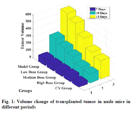
As shown in fig. 1, with the continuous administration, the tumor volume in nude mice in each group continued to increase, but the tumor in the model group doubled and its growth rate was significantly faster than that in the drug group. The tumor volume in the model group was 617.09 mm3 and that in the low dose group was 300.15 mm3, which was much smaller than that in the model group. However, the tumor volume in the high dose group was 553.57 mm3 and that in the medium dose group was 420.35 mm3, indicating that the growth rate of tumor in the low dose group was slower than that in the high dose and middle dose groups slow. At the same time, the experimental values of cyclophosphamide group were the smallest.
As shown in Table 3, during the period from 7 to 13 d after the start of the experiment, the Kinase insert domain receptor (KDR) value in vivo of nude mice in the model group was 0.48, while that in the low-dose group was only 0.35. The KDR value in the low dose group was lower than that in the model group and the difference was statistically significant. There was no significant difference in KDR between low dose group and cyclophosphamide group (0.34).
| Groups | KDR value |
|---|---|
| Model group | 0.48±0.12 |
| High dose group | 0.45±0.15 |
| Medium dose group | 0.41±0.15 |
| Low dose group | 0.35±0.13 |
| Cyclophosphamide group | 0.34±0.10 |
Table 3: Kdr Values in Nude Mice on 7-13 D
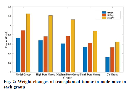
As shown in fig. 2, at the end of the experiment on 13th d, the tumor weight in nude mice in the high dose drug group was 1.21 g, in the medium dose group was 1.12 g, that in the low dose group was 0.88 g and that in the cyclophosphamide group was 0.65 g. The tumor inhibition rates in each group were 3 %, 10 %, 30 % and 48 %, respectively. There was no significant difference in the weight of BGC-823 xenografts between the medium dose and low dose groups (p>0.05), suggesting that the low dose drug group can inhibit the development of BGC-823 tumor. Although the tumor inhibition rate was lower than that of cyclophosphamide group, there was no significant difference between low dose group and cyclophosphamide group (p>0.05).
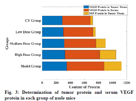
As shown in fig. 3, the expression of VEGF protein in tumor tissue of low dose drug group decreased, only 299.76 mg, while the VEGF protein content in serum of nude mice was 420.32 mg, which was more significant than that in model group (p<0.01). At the same time, VEGF protein content in tumor tissue of high dose drug group was 312.52 mg and that of high dose drug group was 325.46 mg. The results of enzyme linked immunosorbent assay (ELISA) showed that HIF protein expression in tumor tissue of low dose drug group was significantly lower than that of model group, which was only 35.93 mg and the difference was statistically significant (p<0.01).
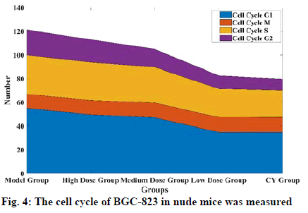
As shown in fig. 4, the percentage of G0 phase cells, G1 phase cells and S phase cells in the low dose group were 34.76 %, 22.76 % and 22.76 %, respectively. The results of double staining showed that the percentage of early apoptosis was 1.97 % in high dose group and 2.18 % in medium dose group, 48 % in the low dose group and 0.44 % in the model group (p<0.01).
HSYA is a kind of blood activating component extracted from cantharus tinctures. In this study, HSYA was used for the first time in nude mice to observe its effect on tumor angiogenesis. Because the tumor inhibition rate of the high dose group in the experiment is too low, the follow up research is not significant, so the follow up gene and protein research are mainly to explore the low dose group. Through the observation results, it can be seen that large, medium and small doses of the drug can delay the growth of transplanted tumor to a certain extent and the effect of HSYA in low dose is the most obvious.
The results showed that VEGF and KDR protein were secreted in the model group and the low dose drug group and VEGF protein in the serum of nude mice were secreted. The expression of VEGF and phosphorylated KDR protein in the low dose group of HSYA was lower and the expression was decreased, especially the VEGF protein in the serum was significantly reduced, suggesting that the appropriate concentration of hydroxy red anthocyanin could inhibit the secretion of VEGF and KDR in BGC-823 xenograft. It is similar to that of VEGF widely expressed in cancer cells of patients with gastric cancer and axon tan sandier decoction can reduce the expression of angiogenic factor gene in orthotopic transplantation of human gastric cancer.
The activation of HIF in eukaryotes can induce the expression of many genes that help the body adapt to hypoxia. The results showed that the level of hydroxy group in HIF group was significantly lower than that in HIF group, which showed that saffron yellow could inhibit the expression of hydroxy group. Hematoxylin and Eosin staining showed that HSYA decreased the proliferation of tumor cells and inhibited tumor growth. Cell apoptosis was obvious, indicating that HSYA may not affect the tumor by affecting cell cycle.
Author’s contributions:
Xiuhua Mao and Zhen Fang have contributed equally to this work as co-first author.
Acknowledgements:
None
Conflict of Interests:
The authors declared no conflict of interest.
References
- Liu L, Si N, Ma Y, Ge D, Yu X, Fan A, et al. Hydroxysafflor-yellow A induces human gastric carcinoma BGC-823 cell apoptosis by activating peroxisome proliferator-activated receptor gamma (PPARγ). Med Sci Monit 2018;24:803.
- Dong XZ, Song Y, Lu YP, Hu Y, Liu P, Zhang L. Sanguinarine inhibits the proliferation of BGC-823 gastric cancer cells via regulating miR-96-5p/miR-29c-3p and the MAPK/JNK signaling pathway. J Nat Med 2019;73(4):777-88.
- Zhang Y, Zhou X, Zhang Q, Zhang Y, Wang X, Cheng L. Involvement of NF-κB signaling pathway in the regulation of PRKAA1-mediated tumorigenesis in gastric cancer. Artif Cells Nanomed Biotechnol 2019;47(1):3677-86.
- Wang T, Duan SJ, Wang SY, Lu Y, Zhu Q, Wang LJ, et al. Coadministration of hydroxysafflor yellow pigment A with levodopa attenuates the dyskinesia. Physiol Behav 2015;147:193-7.
- Ikeguchi M, Cai J, Yamane N, Maeta M, Kaibara N. Clinical significance of spontaneous apoptosis in advanced gastric adenocarcinoma. Cancer 1999;85(11):2329-35.
- Azuma T, Ito S, Sato F, Yamazaki Y, Miyaji H, Ito Y, et al. The role of the HLA?DQA1 gene in resistance to atrophic gastritis and gastric adenocarcinoma induced by Helicobacter pylori infection. Cancer 1998;82(6):1013-8.
- Ajani JA, Winter K, Okawara GS, Donohue JH, Pisters PW, Crane CH et al. Phase II trial of preoperative chemoradiation in patients with localized gastric adenocarcinoma (RTOG 9904): quality of combined modality therapy and pathologic response. J Clin Oncol 2006;24:3953-8.
- Li P, Zhou X, Sun W, Sheng W, Tu Y, Yu Y, et al. Elemene induces apoptosis of human gastric cancer cell line BGC-823 via extracellular signal-regulated kinase (ERK) 1/2 signaling pathway. Med Sci Monit 2017;23:809-17.
- Zhang JF, Sun ZS, Zhang QF, Ding WF, Wu XH, Mao ZB. Expression of long noncoding RNA STCAT3 in gastric cancer tissues and its effect on malignant phenotype of gastric cancer cells. Z Honghua Med J 2016;96(46):3735-40.
- Zhou Y, Li R, Yu H, Wang R, Shen Z. microRNA-130a is an oncomir suppressing the expression of CRMP4 in gastric cancer. Onco Targets Ther 2017;10:3893.
- Zaree A, Javadi HR, Adelipor M, Hojati Z, Kamali M, Bahadoran H. The Study of Anti Genotoxic Effects of Saffron Aqueous Extract in Cadmium Chloride Exposed Mice Kidney by Comet Assay. J Med Plants 2015;14:30-40.
- Soeda S, Aritake K, Urade Y, Sato H, Shoyama Y. Neuroprotective activities of saffron and crocin. Adv Neurobiol 2016;12:275-92.
- Xi S, Zhang Q, Liu, C, Xie H, Yue L. Effects of safflower component HSYA on VEGF Protein, KDR and hypoxia inducible factor expression in human gastric adenocarcinoma BGC-823 Xenograft in Nude Mice. Chin J Tradit Chin Med 2012; 5(11):82-7.
- He L. Study on Bioactive Components of Safflower Extract. Doctoral Dissertation 2016;2(5):21-4.
- Zhang H, Zhang L. Antioxidant Activity of Safflower Yellow Pigment. Chem Res Appl 2012;12(5):715-21.
- Jilibin Z. Effect of Tibetan medicine Xuelingzhi extract on the proliferation of gastric adenocarcinoma cell line MGC-803. Qinghai Univ 2014;3(2):17-9.
- Pan L. The value of spectral CT imaging in the diagnosis of gastric adenocarcinoma and the evaluation of neoadjuvant chemotherapy. Doctoral dissertation 2016;8(12):37-41.


