- *Corresponding Author:
- B. S. Arora
Indian Institute of Integrative Medicine, Jammu Tawi-180 001, India
E-mail: bhupichem27@gmail.com
| Date of Submission | 03 October 2013 |
| Date of Revision | 22 February 2015 |
| Date of Acceptance | 29 November 2015 |
| Indian J Pharm Sci 2015;77(6):792-795 |
This is an open access article distributed under the terms of the Creative Commons Attribution-NonCommercial-ShareAlike 3.0 License, which allows others to remix, tweak, and build upon the work non-commercially, as long as the author is credited and the new creations are licensed under the identical terms.
Abstract
The present study was designed to evaluate the in vitro cytotoxic effect of methanol extract of aerial parts including stems, leaves and twigs of Aralia cachemirica and purified continentalic acid isolated from this extract against a panel of human cancer cell lines of varied tissues. Percentage of growth inhibition was evaluated by sulphorhodamine B assay. Purified continentalic acid showed moderate cytotoxicity against all the cell lines used. In contrast, the extract exhibited significant concentration dependant cytotoxicity against A-549 (lung), THP-1 (leukemia) and MCF-7 (breast) cell lines. This work highlights cytotoxic potential of this extract, which can further be explored for different constituents for their possible use autonomously or in combined manner in cancer therapy. The detailed analysis of their cytotoxicity has been presented in this paper.
Keywords
Aralia cachemirica, continentalic acid, cytotoxicity, sulphorhodamine B assay
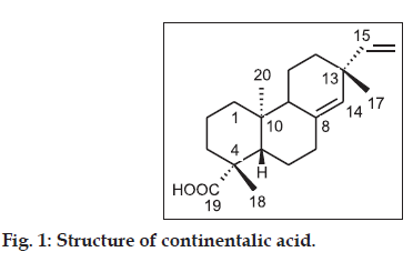
Several species of Aralia (Family Araliaceae) are known for their pharmacological significance [1-7] and have been widely used in traditional medicine for the treatment of rheumatic arthritis, lumbago, lameness [8-10], inflammation [11], gastritis [12], nephritis and diabetes mellitus [13]. Aralia cachemirica is mostly found in India in North Western Himalayas, especially Himachal Pradesh, Uttarakhand and Kashmir [14]. Pharmacological prospective of this plant species has rarely been explored and previous phytochemical and biological studies reported on this species comprise hypoglycemic activity of the roots of Aralia cachemirica [15], isolation of different phytoconstituents including n-tetracont- 19-enoic acid, hexamethyl perhydrophenanthrenyl- 3-ß-n-decanoate, tetrahydrocontinentalic acid, 1ß, 4a, 4ß-trimethyl-6-(10, 14, 18-trimethyl-tridec- 6-enyl)-cyclohexane-4ß-ol, continentalic acid, maltose and sucrose [16], isolation of aralosides and acids [17] and isolation of essential oil from roots of A. cachemirica [18,19]. Isolation of continentalic acid from Aralia cachemirica and evaluation of its immunomodulatory activity has already been reported by our research group [20]. Continentalic acid has been reported for its analgesic activity [21], growth inhibition and apoptosis induction [22], antibacterial activity [23] and antiinflammatory activity [24-26]. Earlier, continentalic acid has been reported to show moderate cytotoxicity against L1210, K562 and LLC tumor cell lines using MTT assay [26]. So far, the cytotoxicity of Aralia cachemirica has not been reported. Herein, we report the evaluation of in vitro cytotoxic effect of crude methanol extract from aerial parts of Aralia cachemirica and purified continentalic acid (CA, fig. 1) isolated from this extract against a panel of five human cancer cell lines by sulphorhodamine B assay to explore their possible role in cancer therapy.
TLC was performed on 0.25 mm silica gel 60 F254 plates. Silica gel 60-120 mesh was used for column chromatography. HPLC analysis was performed using Agilent 1100 series. LC conditions employed were as follows: C8 column reversed-phase (E-Merck, 250×4.0 mm, 5 µm particle size) maintained at 30o, quaternary pump, photodiode array detector, water-acetonitrile (1:4, v/v) isocratic mobile phase at a flow rate of 0.5 ml/min using automatic sample injection module.
The aerial parts of plant Aralia cachemirica were collected from khillanmarg area of Kashmir (Jammu and Kashmir), India after proper identification and authentication of plant by department of Bioscience and Biotechnology, Banasthali University, Rajasthan, India. (Sample no. BV 168). Reference specimen has been preserved in our laboratory. These plant parts were dried in shade and pulverized separately in a mechanical grinder, passed through a 40 mesh sieve, and stored in a closed vessel. This crushed material (680 gms) was extracted with pure methanol through percolator for 72 h. The suspension was filtered and the filtrate was concentrated under reduced pressure to yield 87.95 gm of methanol free semisolid mass. Column chromatography of this extract using n-hexane:ethyl acetate gradient as eluting solution yielded pure continentalic acid as reported earlier [20].
The human cancer cell lines used in present study were procured from National Cancer Institute, Frederick, U.S.A. Cells were grown in tissue culture flasks in complete growth medium (RPMI-1640 medium with 2 mM glutamine, 100 µg/ml streptomycin, pH 7.2, sterilized by filtration and supplemented with 10% fetal calf serum and 100 units/ml penicillin before use) at 37º in an atmosphere of 5% CO2 and 90% relative humidity in a carbon dioxide incubator. These cells at sub confluent stage, when the cells were 60-70% confluent, were harvested from the flask by treatment with trypsin (0.5% in PBS containing 0.02% EDTA). Cells with viability of more than 98%, as determined by trypan blue exclusion, were used for the present study. The cell suspension of the required cell density (1×10⁵ cells/ml) was prepared in complete growth medium containing gentamicin (50 µg/ml) for determination of cytotoxicity. Stock solutions of 4×10⁻² M of continentalic acid and 1000 µg/ml of extract were prepared in DMSO. DMSO was used in such a way that final concentration in the dilution was less than 1%. However the control cells, which were not given any treatment, were having equivalent amounts of DMSO, to nullify the effect. The stock solutions were serially diluted with complete growth medium containing 50 µg/ml of gentamicin to obtain working test solutions of required concentrations and were stored at -20° until use.
In vitro cytotoxicity of test materials was evaluated against the selected human cancer cell lines according to the standard procedure using the protein-binding dye sulforhodamine B to estimate cell growth [27]. The 100 µl of cell suspension was added to each well of the 96-well tissue culture plate. The cells were incubated for 24 h at 37o in an atmosphere of 5% CO2 and 90% relative humidity in a carbon dioxide incubator to allow cell attachment. Test materials of varying concentrations in complete growth medium (100 µl) were added after 24 h incubation to the wells containing cells. The plates were further incubated for 48 h in a carbon dioxide incubator after addition of test material and then the cell growth was stopped by gently layering chilled trichloroacetic acid (50 µl, 50%) on top of the medium in all the wells. The plates were incubated at 4o for one hour to fix the cells attached to the bottom of the wells. The liquid of all the wells was gently decanted and discarded. The plates were washed five times with distilled water and air-dried. Cell growth was measured by staining with sulforhodamine B dye (0.4% w/v in 1% acetic acid) [28]. The adsorbed dye was dissolved in Tris-Buffer (100 ml, 0.01M, pH 10.4) and plates were gently stirred for 5 min on a mechanical stirrer. The optical density (OD) was recorded on ELISA reader at 540 nm. All the assays were done in quadruplets, i.e. four replicates were evaluated.
The cell growth was calculated by subtracting mean OD value of respective blank from the mean OD value of experimental set. Percent growth in presence of test materials was calculated considering the growth in absence of any test material as 100% and in turn percent growth inhibition in presence of test material was calculated.
The cytotoxic effect of methanol extract from stems, leaves and twigs of Aralia cachemirica and purified continentalic acid (CA) isolated from this extract was evaluated by using human cancer cell lines of five different tissues namely A-549 (Lung), DU-145 (Prostate), THP-1 (Leukemia), IMR-32 (Neuroblastoma) and MCF-7 (Breast) at different concentrations for 48 h. The percentage growth inhibition for continentalic acid at 1 µM concentration varied between 0-27% and with increase in concentration to 100 µM, varied between 15-45%. The percentage growth inhibition for the extract at 30 µg/ml varied between 0-51%, but as we increased the concentration to 100 µg/ml, activity also increased and varied between 50-94%. Thus activity was concentration dependant and specific for cell line to cell line. A comparison of percent growth inhibition for the tested materials with the standards has been provided in Table 1.
| Treatment | Concentration | Percentage of growth inhibition* | |||||
|---|---|---|---|---|---|---|---|
| Lung A-549 | Prostate DU-145 | Leukemia THP-1 | Neuroblastoma IMR-32 | Breast MCF-7 | |||
| Extract | 10 mg/ml | 10 | 0 | 9 | 21 | 2 | |
| 30 mg/ml | 27 | 0 | 18 | 51 | 20 | ||
| 100 mg/ml | 79 | 50 | 92 | 66 | 94 | ||
| CA | 1×10⁻⁶ M | 7 | 10 | 11 | 27 | 0 | |
| 1×10⁻⁵ M | 10 | 14 | 13 | 5 | 0 | ||
| 1×10⁻⁴ M | 45 | 20 | 15 | 28 | 38 | ||
| Paclitaxel | 1×10⁻⁵ M | 61 | - | - | - | - | |
| Mitomycin C | 1×10⁻⁵ M | - | 58 | - | - | - | |
| 5-fluorouracil | 2×10⁻⁵ M | - | - | 72 | - | - | |
| Adriamycin | 1×10⁻⁶ M | - | - | 85 | 76 | ||
*As the values denote the mean growth inhibitions of four readings, standard deviation of the values was not feasible. The methanol extract of Aralia cachemirica exhibited concentration dependant activity against the cell lines used for present study, CA: continentalic acid
Table 1: Cytotoxic Effect Of Methanol Extract Of Aerial Parts Of Aralia cachemirica And Purified Continentalic Acid
As shown by the percentage growth inhibition values (Table 1), continentalic acid showed just moderate activity against all cell lines used in the present study, while the extract exhibited significant concentration dependant cytotoxic effect against some specific cell lines, which was comparable to that of the standard cytotoxic agents used for the present study. The concentration of continentalic acid tested against the human cancer cell lines was not increased, because as per the standard protocol, it is always advisable to test any sample in concentrations less than 1x10⁻⁴ M, hence we did not test CA at higher concentrations. The effect exhibited by the extract may be ascribed to some single constituent or to several constituents, or to the combined activity of a number of constituents of the extract. These results indicate that this extract contains other active molecules apart from continentalic acid, which need to be investigated. Hence, this extract has the prospective for further investigation to explore its <em>Aralia cachemirica</em> antitumor activity and to identify its active constituents.
Acknowledgments
Authors are thankful to Dr. R. K. Khajuria and Mr. Rajneesh Anand, Instrumentation division, IIIM (CSIR), Jammu, India for their support in instrumental analysis.
Financial support and sponsorship
Nil.
Conflicts of interest
There are no conflicts of interest.
References
- Lee DH, Seo BR, Kim HY, Gum GC, Yu HH, You HK, et al. Inhibitory effect of Aralia continentalis on the cariogenic properties of Streptococcus mutans. J Ethnopharmacol 2011;137:979-84.
- Oh HL, Lim H, Cho YH, Koh HC, Kim H, Lim Y, et al. HY251, a novel cell cycle inhibitor isolated from Aralia continentalis, induces G1 phase arrest via p53-dependent pathway in HeLa cells. Bioorg Med ChemLett 2009;19:959-61.
- Seo CS, Li G, Kim CH, Lee CS, Jahng Y, Chang HW, et al. Cytotoxic and DNA topoisomerases I and II inhibitory constituents from the roots of Aralia cordata. Arch Pharm Res 2007;30:1404-9.
- Wang J, Li Q, Ivanochko G, Huang Y. Anticancer effect of extracts from a North American medicinal plant – Wild sarsaparilla. Anticancer Res 2006;26:2157-64.
- Chung YS, Choi YH, Lee SJ, Choi SA, Lee JH, Kim H, et al. Water extract of Aralia elata prevents cataractogenesis in vitro and in vivo. J Ethnopharmacol 2005;101:49-54.
- Park HJ, Hong MS, Lee JS, Leem KH, Kim CJ, Kim JW, et al. Effects of Aralia continentalis on hyperalgesia with peripheral inflammation. Phytother Res 2005;19:511-3.
- Ryu SY, Ahn JW, Han YN, Han BH, Kim SH. In vitro antitumor activity of diterpenes from Aralia cordata. Arch Pharm Res 1996;19:77-8.
- Lim TY. Comparative study on the pharmacological effect of kinds of Dokhwal. J Korean Orient Med 1987;8:97-8.
- Kim JS, Kang SS. Saponins from the aerial parts of Aralia continentalis. Nat Prod Sci 1998;4:45-50.
- Yoshihara K, Hirose Y. Terpenes from Aralia species. Phytochemistry 1973;12:468.
- Han BH, Woo ER, Park MH, Han YN. Studies on the anti-inflammatory activity of Aralia continentalis (III). Arch Pharm Res 1985;8:59-65.
- Lee EB, Kim OJ, Kang SS, Jeong C. Araloside A, an antiulcer constituent from the root bark of Aralia elata. Biol Pharm Bull 2005;28:523-6.
- Yoshikawa M, Murakami T, Harada E, Murakami N, Yamahara J, Matsuda H. Bioactive saponins and glycosides. VII. On the hypoglycemic principles from the root cortex of Aralia elata seem: Structure related hypoglycemic activity of oleanolic acid oligoglycoside. Chem Pharm Bull (Tokyo) 1996;44:1923-7.
- Pusalkar PK. A new species of Aralia [Araliaceae, Sect.: Pentapanax (Seem.) J. Wen] from Jammu and Kashmir, North-west Himalaya, India. Taiwania 2009;54:226-30.
- Bhat ZA, Ansari SH, Mukhtar HM, Naved T, Siddiqui JI, Khan NA. Effect of Aralia cachemiricaDecne root extracts on blood glucose level in normal and glucose loaded rats. Pharmazie 2005;60:712-3.
- Bhat Z, Ali M, Ansari SH, Naquvi KJ. New phytoconstituents from the roots of Aralia cachemiricaDecne. J Saudi ChemSoc 2015;19:287-91.
- George V, Nigam SS, Rishi AK. Isolation and characterization of aralosides and acids from Aralia cachemirica. Fitoterapia
- 1984;55:124-6.
- Shawl AS, Bhat KA, Bhat MA. Essential oil composition of Aralia cachemirica. Indian Perfum 2009;53:35-6.
- Verma RS, Padalia RC, Yadav A, Chauhan A. Essential oil composition of Aralia cachemirica from Uttarakhand, India. Rec Nat Prod 2010;4:163-6.
- Sharma E, Arora BS, Khajuria A, Sidiq T, Kishore D, Vishwakarma RA. Isolation of continentalic acid from Aralia cachemirica and itsimmuno-biological evaluation. Int J Pharm Sci Res 2011;2:2183-9.
- Okuyama E, Nishimura S, Yamazaki M. Analgesic principles from Aralia cordataThunb. Chem Pharm Bull (Tokyo) 1991;39:405-7.
- Kwon TO, Jeong SI, Kwon JW, Kim YC, Jang SI. Continentalic acid from Aralia continentalis induces growth inhibition and apoptosis in HepG2 cells. Arch Pharm Res 2008;31:1172-8.
- Jeong SI, Han WS, Yun YH, Kim KJ. Continentalic acid from Aralia continentalis shows activity against methicillin-resistant Staphylococcus aureus. Phytother Res 2006;20:511-4.
- Han BH, Han YN, Han KA, Park MH, Lee EO. Studies on the anti-inflammatory activity of Aralia continentalis (I). Arch Pharm Res 2006;6:17-23.
- Dang NH, Zhang X, Zheng M, Son KH, Chang HW, Kim HP, et al. Inhibitory constituents against cyclooxygenases from Aralia cordataThunb. Arch Pharm Res 2005;28:28-33.
- Lee IS, Jin W, Zhang X, Hung TM, Song KS, Seong YH, et al. Cytotoxic and COX-2 inhibitory constituents from the aerial parts of Aralia cordata. Arch Pharm Res 2006;29:548-55.
- Monks A, Scudiero D, Skehan P, Shoemaker R, Paull K, Vistica D, et al. Feasibility of a high-flux anticancer drug screen using a diverse panel of cultured human tumor cell lines. J Natl Cancer Inst 1991;83:757-66.
- Skehan P, Storeng R, Scudiero D, Monks A, McMahon J, Vistica D, et al. New colorimetric cytotoxicity assay for anticancer-drug screening. J Natl Cancer Inst 1990;82:1107-12.
