- Corresponding Author:
- H. Kang
Department of Medical Laboratory Science, College of Health Science, Dankook University, Cheonan, 330-714
E-mail: hkang@dankook.ac.kr
| Date of Submission | 23 October 2013 |
| Date of Revision | 22 April 2014 |
| Date of Acceptance | 27 April 2014 |
| Indian J Pharm Sci 2014; 76(4): 267-273 |
Abstract
Houttuynia cordata Thunb (Saururaceae) is a traditional medicinal herb used to treat several disease symptoms. The present study was focused on the hepatoprotective effects of H. cordata ethyl acetate extract in experimental mice. Further the antioxidant potential of the extract was also evaluated to substantiate its hepatoprotective properties. Carbon tetrachloride-induced hepatic damage in mice was used to measure the serum biochemical parameters. Morphological changes in hepatocyte architecture were studied by haematoxylin and eosin staining. In vitro alkyl and hydroxyl free radical scavenging assays were performed to evaluate the antioxidant effect. Administration of H. cordata extract significantly reduced the elevated serum levels and regulated the altered levels of serum cholesterol in carbon tetrachloride-treated mice (P<0.05). The morphological changes in hepatocyte architecture were also reversed by H. cordata treatment. Further, the extract showed significant antioxidant actions by scavenging the alkyl and hydroxyl free radicals. The concentration of the extract necessary for 50% scavenging of alkyl and hydroxyl radicals was 15.5 and 410 µg/ml, respectively. H. cordata extract exhibited significant hepatoprotective property in carbon tetrachloride-induced hepatotoxicity in mice. The strong antioxidant activities possessed by the extract might be responsible for such actions.
Keywords
Heartleaf, antioxidant, hydroxyl radicals, hepatotoxicity, hyperlipidemia, carbon tetrachloride
Liver is an important organ which plays a central role in metabolic homeostasis [1]. Hepatitis, environmental pollutants, bacterial and viral infections, alcohol abuse are said to be some of the major factors leading to liver failure. In recent decades, the pharmaceutical application potential of natural products from herbs and dietary supplement has attracted much interest from researchers in treating liver complications [2,3]. In experimental hepatopathy, carbon tetrachloride (CCl4) has been commonly used because it initiates oxidative damage, decreases the activities of antioxidant enzymes and generates toxic free radicals [4,5]. CCl4- induced hepatotoxicity in animal models has been shown to be similar to human liver cirrhosis [6,7].
Antioxidants of plant origin have been demonstrated to either inhibit or prevent the progression of cellular disturbances resulting from the CCl4-induced liver injury [8]. Houttuynia cordata Thunb (Saururaceae, H. cordata) is a perennial herb that is native to Southeast Asia. It is known as Eo-Sung-Cho in Korea and elaborates a wide spectrum of medicinal properties including allergic inflammation and anaphylaxis [9,10]. Traditionally, H. cordata was used as folk remedy for diuresis, antiviral, antibacterial and antileukemic activities [11]. However, the hepatoprotective protective actions and effect on serum lipid profiles of H. cordata in CCl4-induced hepatotoxicity in experimental animals have not been addressed. In this study, H. cordata ethyl acetate extract was evaluated for its hepatoprotective effects using CCl4-induced acute hepatic damage in experimental mice. Further the in vitro antioxidant activities of H. cordata extract has also been investigated.
One of the major features of metabolic syndrome is dyslipidemia, characterized by elevated levels of very low-density lipoprotein (VLDL), triglyceride (TG) and low density lipoprotein (LDL) [12]. Elevated plasma VLDL and TG levels are due, in large part, to an increase in hepatic overproduction of TG-enriched VLDL particles [13]. CCl4-induced toxicity, which is thoroughly studied for its hepatotoxic properties, also in turn increases the accumulation of fat in the liver leading to hyperlipidemia. Mounting evidence suggests that reactive oxygen species (ROS) are known to play a pivotal role in liverdisease pathology and ROS have been proven to be associated with CCl4-induced hepatotoxicity [14,15]. Therefore, antioxidant therapy might be an ideal approach to ameliorate liver damage caused by hepatotoxin.
Materials and Methods
Male ICR mice (15-16 g) were obtained from NARA Biotech (Seoul, Korea). The animals were kept under standard laboratory conditions. A 12 h light/dark cycle in a temperature and humidity controlled room with water and food ad libitum was maintained. All animal experiments were performed in accordance and approval with our Institutional Animal Care and Use Committee of Konkuk University and the International Guidelines for Handling of Laboratory Animals [16]. Mice were anaesthetized by ketamine (160 mg/kg)/xylazine (40 mg/kg) cocktail injections, intraperitoneally.
Preparation of the H. cordata extract
Dried plant material of H. cordata was purchased from the traditional herb market and was authenticated by a taxonomist at Konkuk University, South Korea. A voucher specimen (HC-KU2013) was kept in our department herbarium for future reference. To obtain the H. cordata extract, the dried plant material was ground in a mixer and defatted three times with three volumes of ethanol. The residue was extracted with absolute ethanol at 1:10 ratio (w/v) for 2 h in heating mantle at 70~80°. The supernatant was filtered and concentrated in a rotary evaporator at 50°. For further fractionation, the alcoholic extract (50 g) was partitioned into hexane, ethyl acetate (EA), and n-butanol fractions to yield 0.53, 8.72 and 38.25 g, respectively. The EA fraction of H. cordata (HCE) was redissolved in distilled water and used for evaluating hepatoprotective activities. Acute toxicity studies on HCE extract was performed at various doses (50, 100, 200 and 400 mg/kg) and was found that upto 400 mg/kg of HCE did not show any signs of toxicity in mice (data not shown). All reagents used in this study were of highest grade available commercially.
Carbon tetrachloride-induced hepatic injury
The animals were randomly divided into four groups of six mice each. Group A served as the normal vehicle control and was administrated orally with 2 ml/kg corn oil daily for a period of 7 weeks. Group B orally administrated with 2 ml/kg body weight of CCl4 (20% CCl4 in corn oil) once a day for 7 weeks. The animals of Group C were pretreated with silymarin (200 mg/kg per day, dissolved into 0.1% DMSO) served as positive control and group D were pretreated with HCE extract (200 mg/kg per day) orally for 7 weeks, respectively before CCl4 treatment. At the end of experiment, animals were sacrificed and the blood samples were collected into heparinized tubes for each group separately. Liver tissue was dissected out, washed immediately with ice cold saline, weighted and then immediately stored at -70° until analysis.
Biochemical assessment
Liver damage was assessed by the estimation of serum activities of ALT, ALP and AST using commercially available test kits from BioVision (Milpitas, CA, USA) according to manufacturer?s instructions. Total cholesterol, HDL-cholesterol, LDL/ VLDL-cholesterol, TG levels were also estimated by using the respective diagnostic kits from BioVision.
Haematoxylin and eosin (H&E) staining
A portion of the left lobe of the liver was preserved in 10% neutral formalin solution for at least 24 h, processed and paraffin embedded as per the standard protocol. Sections of 5 mm in thickness were cut, deparaffinised, dehydrated, and stained with H&E for the estimation of hepatocyte architecture.
Alkyl radical scavenging assay
Alkyl radicals were generated by 2,2-azobis (2-amidinopropane) hydrochloride (AAPH, Sigma Chemical Co., St. Louis, MO, USA). Reaction mixtures containing 40 mM AAPH, 40 mM 4-POBN, and the indicated concentrations of tested samples diluted in PBS (pH 7.4) were incubated at 37° in a water bath for 30 min [17] and then transferred to 50 µl Teflon capillary tubes. The final concentration of HCE was 100 µg/ml. The spin adduct was recorded using the ESR spectrometer. The following measurement conditions were used: Central field 3475 G, modulation frequency 100 kHz, modulation amplitude 2 G, microwave power 10 mW, gain 6.3×10⁵, and temperature 298 K. The radical-scavenging activity of the HCE at various concentrations was calculated according to the following formula: Scavenging rate=(h0-hx)/h0×100%, where h0 and hx are the ESR signal intensities of samples with and without extract, respectively.
Hydroxyl radical scavenging assay
Hydroxyl radicals were generated by the Fenton reaction and reacted rapidly with nitrone spin trap 5,5-dimethyl-1-pyrroline N-oxide (DMPO, Sigma Chemical Co., St. Louis, MO, USA). The resulting DMPO-OH adduct was detected by ESR [18]. The ESR spectrum was recorded 2.5 min after mixing with phosphate buffer solution (pH 7.4) supplemented with 20 µl of 0.3 M DMPO, 20 µl of 10 mM FeSO4, and 20 µl of 10 mM H2O2 under the following conditions: Central field 3475 G, modulation frequency 100 kHz, modulation amplitude 2 G, microwave power 1 mW, gain 6.3×10⁵, and temperature 298 K. The radical-scavenging activity of the HCE at various concentrations was calculated according to the formula mentioned as in alkyl radical scavenging assay.
Statistical analysis
All data are represented as the mean±SEM of at least three independent experiments. Statistical analyses were performed using SAS statistical software (SAS Institute, Cray, NC, USA) using Student?s t-test and one-way analysis of variance, followed by Dunnett?s multiple range tests. In all experiments P values less than 0.05 was considered statistically significant.
Results
Effect of HCE extract on CCl4-induced hepatotoxicity
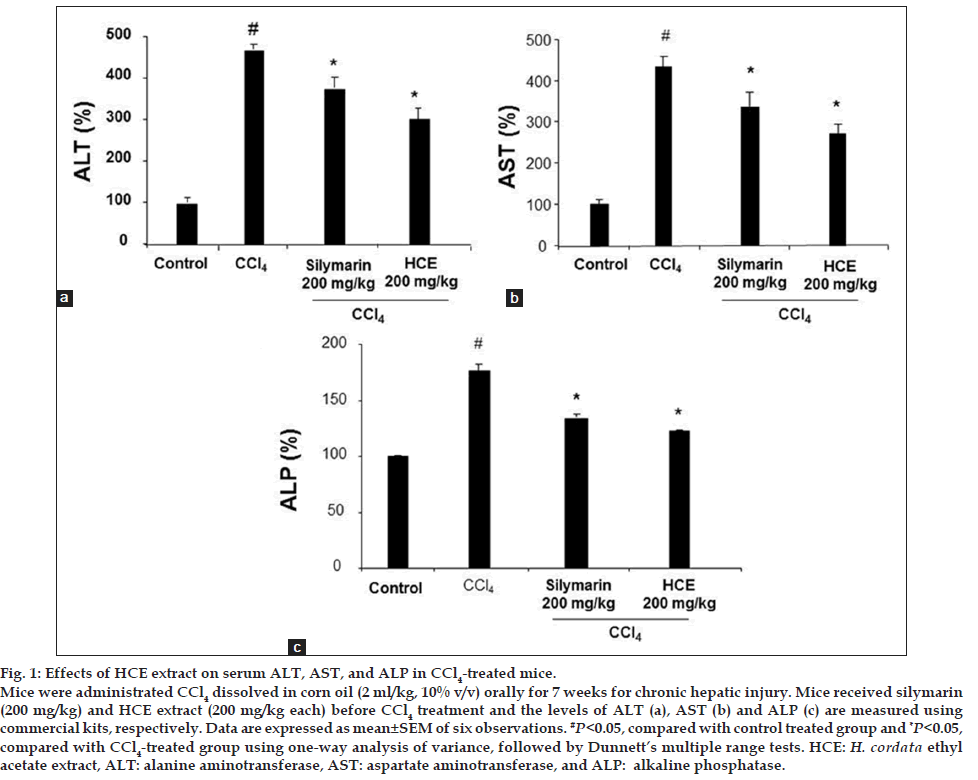
The serum biochemical data for the evaluation of CCl4- induced hepatotoxicity by measuring the ALT and AST as conventional indicator of the liver injury are shown in fig. 1a-c. There was a remarkable elevation of serum ALT, AST and ALP activities in the CCl4-treated group as compared to the vehicle control (nearly, 5 fold for ALT (P<0.05), 4.5 fold for AST (P<0.05) and 1.8 fold for ALP (P<0.05), respectively indicating CCl4-induced damage to the hepatic cells. However, administration of HCE extract at a dosage of 200 mg/kg suppressed the elevated serum ALT, AST and ALP in a significant manner when compared to CCl4 alone treated group (P<0.05). The positive control silymarin treated group (200 mg/kg) also exhibited significant attenuation (P<0.05) when compared to CCl4 treated groups. These results demonstrate that the levels of ALT, AST and ALP were significantly restored by treatment with HCE extract and the attenuation is on par with standard drug silymarin treated groups.
Figure 1: Effects of HCE extract on serum ALT, AST, and ALP in CCl4-treated mice.
Mice were administrated CCl4 dissolved in corn oil (2 ml/kg, 10% v/v) orally for 7 weeks for chronic hepatic injury. Mice received silymarin
(200 mg/kg) and HCE extract (200 mg/kg each) before CCl4 treatment and the levels of ALT (a), AST (b) and ALP (c) are measured using
commercial kits, respectively. Data are expressed as mean±SEM of six observations. #P<0.05, compared with control treated group and *P<0.05,
compared with CCl4-treated group using one-way analysis of variance, followed by Dunnett?s multiple range tests. HCE: H. cordata ethyl
acetate extract, ALT: alanine aminotransferase, AST: aspartate aminotransferase, and ALP: alkaline phosphatase.
Effects of HCE extract on CCl4-treated elevated lipid levels
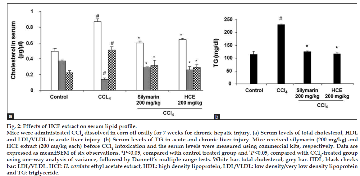
The effect of HCE extract on the cholesterol profile (total cholesterol, LDL/VLDL, HDL and TG) in CCl4-induced liver injury in mice was shown in fig. 2a and b. A significant increase in the total cholesterol, LDL/VLDL and TG levels were observed in CCl4-treated mice, compared to the control group (P<0.05). In contrast the HDL levels were decreased significantly in CCl4-treated group. However, pretreatment with HCE extract (200 mg/kg) significantly attenuated the CCl4-induced increased serum levels of total cholesterol, LDL/VLDL and TG levels (P<0.05) indicating that HCE extract showed efficacy to possess antihyperlipidemic activity. Further, the CCl4-induced decrease in HDL serum levels were reversed significantly to near normal conditions when pretreated with HCE extract (P<0.05). Silymarin (200 mg/kg) also attenuated the increased levels of total cholesterol, LDL/VLDL and TG and the decreased level of HDL, which was consistent with the results from other researchers (P<0.05) [19].
Figure 2: Effects of HCE extract on serum lipid profile.
Mice were administrated CCl4 dissolved in corn oil orally for 7 weeks for chronic hepatic injury. (a) Serum levels of total cholesterol, HDL
and LDL/VLDL in acute liver injury. (b) Serum levels of TG in acute and chronic liver injury. Mice received silymarin (200 mg/kg) and
HCE extract (200 mg/kg each) before CCl4 intoxication and the serum levels were measured using commercial kits, respectively. Data are
expressed as mean±SEM of six observations. #P<0.05, compared with control treated group and *P<0.05, compared with CCl4-treated group
using one-way analysis of variance, followed by Dunnett?s multiple range tests. White bar: total cholesterol, grey bar: HDL, black checks
bar: LDL/VLDL. HCE: H. cordata ethyl acetate extract, HDL: high density lipoprotein, LDL/VLDL: low density/very low density lipoprotein
and TG: triglyceride.
Effects of HCE on CCl4-induced hepatic morphological changes
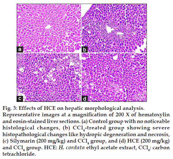
The protective effect exerted by HCE extract against CCl4-induced hepatotoxicity was further confirmed by conventional histological assessment (fig. 3). As shown in fig. 3a, the histology of the liver sections of control group showed normal hepatic cells. The stained sections of CCl4 model group revealed extensive liver injuries characterized by moderate to severe hepatocellular hydropic degeneration and necrosis (fig. 3b). However, CCl4-treated mice pretreated with 200 mg/kg silymarin or HCE extract (200 mg/kg) markedly ameliorated the hepatic lesions (fig. 3c and d), respectively, when compared with CCl4 alone treated groups.
Figure 3: Effects of HCE on hepatic morphological analysis.
Representative images at a magnification of 200 X of hematoxylin
and eosin-stained liver sections. (a) Control group with no noticeable
histological changes, (b) CCl4-treated group showing severe
histopathological changes like hydropic degeneration and necrosis,
(c) Silymarin (200 mg/kg) and CCl4 group, and (d) HCE (200 mg/kg)
and CCl4 group. HCE: H. cordata ethyl acetate extract, CCl4: carbon
tetrachloride.
Alkyl free radical scavenging effects of HCE extract
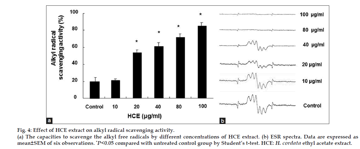
The alkyl free radical scavenging activity of the HCE extract was shown in fig. 4. The alkyl radical spin adduct of 4-POBN/free radicals was generated from AAPH at 37° after 30 min, and a decrease in the ESR signals was observed at increasing HCE concentrations. The alkyl radical scavenging activity was 90.5% at 100 µg/ml (fig. 4a), and the IC50 value of the HCE extract was 15.50 µg/ml. The decrease in the ESR signals for alkyl radical scavenging effect of HCE was shown in fig. 4b.
Figure 4: Effect of HCE extract on alkyl radical scavenging activity.
(a) The capacities to scavenge the alkyl free radicals by different concentrations of HCE extract. (b) ESR spectra. Data are expressed as
mean±SEM of six observations. *P<0.05 compared with untreated control group by Student?s t-test. HCE: H. cordata ethyl acetate extract.
Hydroxyl radical scavenging effects of HCE extract
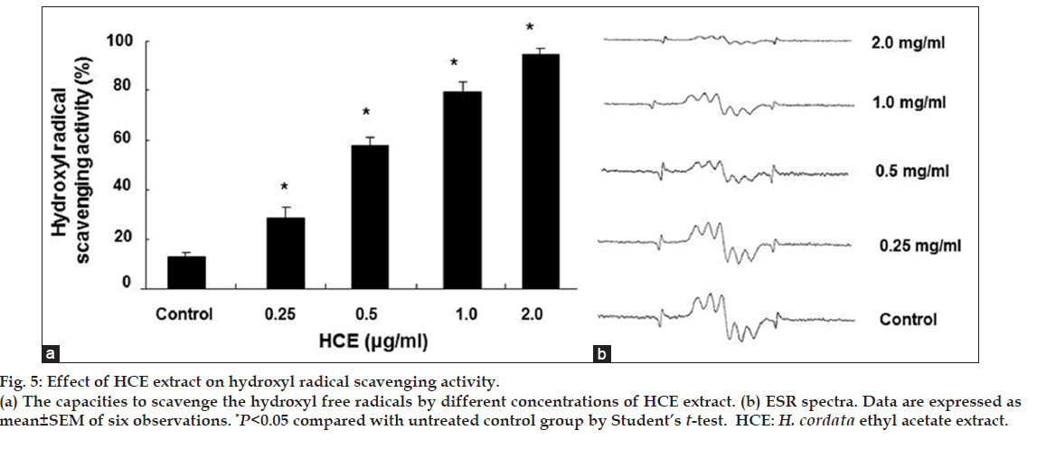
The hydroxyl radical generated in the Fe2+/H2O2 system was trapped by DMPO, which formed the spin adduct detected by ESR spectroscopy, and the typical 1:2:2:1 ESR signal of the DMPO-OH adduct was observed. The height of the third peak in the spectrum represents the relative amounts of DMPO-OH adduct. The addition of the HCE extract resulted in a decrease in the peak corresponding to the DMPO-OH adducts. The maximum hydroxyl radical scavenging effect of HCE was found to be 87.6% at 2 mg/ml and the IC50 value of HCE was determined to be 410 µg/ml (fig. 5a). The decrease in the ESR signals for alkyl radical scavenging effect of HCE was shown in fig. 5b. These results clearly demonstrate that the HCE possesses antioxidative activity and can effectively scavenge alkyl and hydroxyl radicals.
Figure 5: Effect of HCE extract on hydroxyl radical scavenging activity.
(a) The capacities to scavenge the hydroxyl free radicals by different concentrations of HCE extract. (b) ESR spectra. Data are expressed as
mean±SEM of six observations. *P<0.05 compared with untreated control group by Student?s t-test. HCE: H. cordata ethyl acetate extract.
Discussion
This study was undertaken to demonstrate the protective ability of HCE extract against liver damage induced by CCl4 in mice. Mounting evidence suggests that the elementary lesions caused by CCl4-treatment in experimental animals are similar to those seen in most cases of human liver diseases [6,7]. Therefore this model has been used for decades to investigate and screen various natural and synthetic agents in the prevention and/or treatment of liver injuries. It was well documented that CCl4 administration produce toxic free radicals thereby causing liver damage which further leads to the elevated levels of liver enzymes and altered levels of serum lipid profile disrupting the liver function [20]. Antioxidant rich natural herbs and their active constituents were well reported to exert beneficial effects in various disease conditions including liver diseases [14,15].
The ALT, AST and ALP, commonly referred as liver enzymes which are released from damaged hepatocytes and their levels in the serum have been widely recognized as a useful quantitative marker to study the extent and type of hepatocellular injuries [21]. In our experiment, HCE extract significantly attenuated the elevated serum levels of ALT, AST and ALP indicating that the proportion of damaged hepatocytes was reduced as a direct result of HCE administration.
Liver plays an important role in the regulation of lipoprotein transport in serum and cholesterol metabolism. During lipoprotein transport, LDL and HDL appear to be especially pivotal. LDL, rich in cholesterol and cholesterol ester, is regarded as bad cholesterol, whereas HDL contains relatively little cholesterol and is regarded as good cholesterol. Reports suggested that high levels of serum cholesterol, LDL, and TG and low levels of HDL were induced with CCl4 in animal model systems [17]. Therefore, to evaluate whether HCE extract has any influence in hyperlipidemia induced by CCl4, cholesterol catabolism was investigated. In the present study HCE extract significantly prevented CCl4- induced liver damage as evidenced by reduced serum concentrations of cholesterol contents. Data form our study indicated that HCE extract restored the lipid levels to near normal levels.
The histopathological analysis provided complementary evidence that pretreatment with HCE extract attenuated the morphological changes in mouse liver tissue induced by CCl4 administration. Examination of liver sections of mice, which received CCl4, revealed disruption of the normal structural organization of hepatocytes compared to normal liver cells. Many hepatic cells were damaged and lost their characteristic appearance. In contrast treatment with HCE extract restored the hepatocellular architecture in CCl4-induced mice.
Silymarin, a natural antioxidant was well reported to restore liver enzyme activities due to its antioxidant properties, chelating metal ions, inhibiting lipid peroxidation, protecting the membrane permeability properties, and preventing liver glutathione depletion in CCl4-induced liver injury in mice [19]. In our present study, HCE extract showed similar or significantly greater effect when compared with the effects of silymarin at the same dose tested in CCl4-induced liver damage in mice, indicating the potential of HCE extract in liver protection.
Earlier studies suggest that H. cordata inhibited lipidperoxidation in rat liver homogenates and possess antioxidant effects [22]. Recent report from our lab also indicated that H. cordata possess antineuroinflammatory activity by virtue of its antioxidant effects [23]. Data from our present work showed that HCE significantly inhibited alkyl and hydroxyl free radicals. Phytochemical studies on H. cordata showed that it contains several polyphenolic agents such as quercitrin, quercetin, rutin, hyperin, isoquercitrin, quercitrin, ß-myrcene, ß-pinene, a-pinene, a-terpineol and n-decanoic acid [24]. The antioxidant polyphenolic compounds present in HCE extract might be responsible for delivering such potent hepatoprotective actions in CCl4-treated mice. In conclusion, our findings showed that HCE extract significantly prevented CCl4-induced liver damage as evidenced by decreased serum activities of AST, ALT and ALP.
HCE extract also modulated serum concentrations of cholesterol contents. Further HCE exhibited potent antioxidant effects substantiating its beneficial effects in hepatoprotection.
References
- Taub R. Liver regeneration: From myth to mechanism. Nat Rev Mol Cell Biol 2004;5:836-47.
- Hinds TS, Wesk WL, Knight EM. Carotenoids and retinoids: A review of research, clinical and public health applications. J Clin pharmacol 1997;37:551-8
- He YT, Liu DW, Ding LY, Li Q, Xiao YH. Therapeutic effects and molecular mechanical of antifibrosis herbs and selection on rats with hepatic fibrosis. World J Gastroenterol 2004;10:703-6.
- Poli G. Liver damage due to free radicals. Br Med Bull 1993;49:604-20.
- Ohta Y, Kongo M, Sasaki E, Nishida K, Ishiguro I. Therapeutic effect of melatonin on carbon tetrachloride-induced acute liver injury in rats. J Pineal Res 2000;28:119-26.
- Parola M, Leonarduzzi G, Biasi F, Albano E, Biocca ME, Poli G, et al. Vitamin E dietary supplementation protects against carbon tetrachloride-induced chronic liver damage and cirrhosis. Hepatology 1992;16:1014-21.
- Thrall KD, Vucelick ME, Gies RA, Zangar RC, Weitz KK, Poet TS, et al. Comparative metabolism of carbon tetrachloride in rats, mice, and hamsters using gas uptake and PBPK modeling. J Toxicol Environ Health A 2000;60:531-48.
- Rajesh MG, Latha MS. Perliminary evaluation of the antihepatotoxic activity of Kamilari, a polyherbal formulation. J Ethnopharmacol 2004;91:99-104.
- Lee JS, Kim IS, Kim JH, Kim JS, Kim DH, Yun CY. Suppressive effects of Houttuynia cordata Thumb (Saururaceae) extract on Th2 immune response. J Ethnophamacol 2008;117:34-40.
- Li GZ, Chai OH, Lee MS, Han EH, Kim HT, Song CH. Inhibitory effects of Houttuynia cordata water extracts on anaphylactic reaction and mast cell activation. Biol Pharm Bull 2005;28:1864-8.
- Lai KC, Chiu YJ, Tang YJ, Lin KL, Chiang JH, Jiang YL, et al. Houttuynia cordata thumb extract inhibits cell growth and induces apoptosis in human primary colorectal cancer cells. Anticancer Res 2010;30:3549-56.
- 12. Moller DE, Kaufman KD. Metabolic syndrome: A clinical and molecular perspective. Annu Rev Med 2005;56:45-62.
- 13. Adiels M, Taskinen MR, Packard C, Caslake MJ, Soro-Paavonen A, Westerbacka J, et al. Overproduction of large VLDL particles is driven by increased liver fat content in man. Diabetologica 2006;49:755-65.
- Recknagel RO, Glende EA Jr, Dolak, JA, Waller RL. Mechanisms of carbon tetrachloride toxicity. Pharmacol Ther 1989;43:139-54.
- Slater TF, Sawyer BC. The stimulatory effects of carbon tetrachloride on peroxidative reactions in rat liver fractions in vitro. Inhibitory effects of free radical scavengers and other agents. Biochem J 1971;123:823-8
- Derrell C. Guide for the care and use of laboratory animals. Institute of Laboratory Animal Resources. Washington DC, USA: National Academy Press; 1996.
- Lin HM, Tseng HC, Wang CJ, Lin JJ, Lo CW, Chou FP. Hepatoprotective effects of Solanum nigrum Linn. Extract against CCl4-induced oxidative damage in rats. Chem Biol Interact 2008;171:283-93.
- Yang YS, Ahn TH, Lee JC, Moon CJ, Kim SH, Jun W, et al. Protective effects of Pycnogenol on carbon tetrachloride-induced hepatotoxicity in Sprague-Dawley rats. Food Chem Toxicol 2008;46:380-7.
- Chen IS, Chen YC, Chou CH, Chuang RF, Sheen LY, Chiu CH. Hepatoprotection of silymarin against thioacetamide-induced chronic liver fibrosis. J Sci Food Agric 2012;92:1441-7.
- Kumar P, Sivaray A, Elumalai E, Kumar B. Carbon tetrachloride? induced hepatotoxicity in rats-protective role of aqueous leaf extracts of Coccinia grandis. Int J Pharm Tech Res 2009;1:1612-5.
- Khan RA, Khan MR, Sahreen S, Shah NA. Hepatoprotective activity of Sonchus asper against carbon tetrachloride-induced injuries in male rats: A randomized controlled trial. BMC Complement Altern Med 2012;12:90.
- Ng LT, Yen FL, Liao CW, Lin CC. Protective effect of Houttuynia cordata extract on Bleomycin-induced pulmonary fibrosis in rats. Am J Chin Med 2007;35:465-75.
- Park TK, Koppula S, Kim MS, Jung SH, Kang H. Anti- Neuroinflammatory effects of Houttuynia cordata extract on LPSstimulated BV-2 microglia. Trop J Pharm Res 2013;12:523-8.
- Nuengchamnong N, Krittasilp K, Ingkaninan K. Rapid screening and identification of antioxidants in aqueous extracts of Houttuynia cordata using LC-ESI-MS coupled with DPPH assay. Food Chem 2009;117:750-6.
