- *Corresponding Author:
- A. H. M. Viswanathaswamy
Department of Pharmacology, K. L. E. S College of Pharmacy, Vidyanagar, Hubli - 580 031, India
E-mail: vmhiremath2004@yahoo.com
| Date of Submission | 24 August 2009 |
| Date of Revision | 23 December 2009 |
| Date of Acceptance | 10 May 2010 |
| Indian J. Pharm. Sci., 2010, 72 (3): 367-371 |
Abstract
The present study was designed to compare ulcer protective effect of proton pump inhibitors viz. omeprazole, rabeprazole and lansoprazole against dexamethasone plus pylorus ligation induced ulcer model. Dexamethasone (5 mg/kg) was used as an ulcerogen. Dexamethasone suspended in 1% CMC in water was given orally to all the rats 15 min after the pylorus ligation. Omeprazole (20 mg/kg), rabeprazole (20 mg/kg), and lansoprazole (20 mg/kg) were administered by oral route 30 min prior to ligation was used for ulcer protective studies, gastric secretion and mucosal studies. Effects of proton pump inhibitors were determined by the evaluation of various biochemical parameters such as ulcer index, free and total acidity, gastric pH, mucin, pepsin and total proteins. Oral administration of proton pump inhibitors showed significant reduction in gastric acid secretion and ulcer protective activity against dexamethasone plus pylorus ligation induced ulcer model. The % protection of omeprazole, rabeprazole and lansoprazole was 84.04, 89.36 and 79.78, respectively. Rabeprazole significantly inhibited the acid-pepsin secretion and increased the gastric mucin secretion. The observations made in the present study suggest that rabeprazole is the most effective gastric antisecretory and ulcer healing agent as compared to omeprazole and lansoprazole.
Keywords
Dexamethasone, PPIs, mucosal offensive and defense factors
Acute gastric ulcers occur due to erosion in the mucosal membrane, generally in gastric and duodenum regions and occur as a result of alteration in the balance between mucosal damaging agents and mucosal defense mechanisms. Peptic ulcer is one of the most frequent disorders of the alimentary tract and in various countries its prevalence is estimated as 5-10% of the adult population [1]. This disorder remains one of the most important problems, both in the practice of primary health care physicians and gastroenterologists. It has been reported earlier that heavy smoking, alcohol and steroids intake may delay healing of ulcers. This could be due to increased gastric acid secretion, reduction in gastric mucosal blood flow, inhibition of duodenal bicarbonate production, prostaglandin synthesis, Helicobacter pylori infection, reduced generation of nitric oxide and increased generation of free radicals [2-5]. Ulcerogenic potential of corticosteroids is well known as a result of increased gastric acid and pepsin secretion, which will aggravate peptic ulcer [6]. Frequent usage of corticosteroids in the treatment of bronchial asthma, brain metastasis, cerebral edema etc has increased the risk of peptic ulcer [7]. Corticosteroids cause reduction in the levels of nitric oxide [8], inhibition of prostaglandin (PG) synthesis and formation of lipid peroxides [9] leading to gastric erosions by damaging surface epithelial cells.
Proton pump inhibitors (PPIs) inhibit release of hydrogen ion from parietal cells. It inhibits gastric acid secretion by blocking H+/K+ ATPase pump. Omeprazole shows an ulcer healing effect by inhibiting neutrophil chemotaxis, superoxide production and release of active oxygen metabolites [10] leading to ulcer healing by augmenting luminal pH there by decreasing pepsin damage to gastric mucosa.
While lansoprazole prevents gastric mucosal damage by gastric prostaglandin production, expression of cyclo-oxygenase (COX) isoforms and release of stable nitric oxide metabolites into gastric juice and blocks the oxygen derived free radical output from neutrophils activated by Helicobacter pylori and exerts its antioxidant effect [11,12].
Rabeprazole causes perhaps the fastest acid suppression and so aid gastric mucin synthesis. This is necessary for the maintenance of mucosal integrity. Although these PPIs being similar in pharmacological actions they differ in clinical pharmacology [13]. Therefore, the present work was undertaken with an aim to compare different PPIs for the treatment of dexamethasone plus pylorus ligation induced ulcer model in albino rats.
Omeprazole was obtained from Cipla Ltd, Goa, India. Rabeprazole, lansoprazole and dexamethasone were obtained from Cadila health care, Ahmedabad, India. The chemicals and solvents used were sodium hydroxide, Topfer’s reagent, copper sulphate, phenolphthalein, sodium carbonate, phenol reagent, bovine albumin, sucrose, alcian blue, sodium acetate, ethanol, methanol, dil.HCl and chloroform. All were of analytical grade.
Healthy Wistar rats of either sex weighing between 150-200 g were used. Animals were housed individually in polypropylene cages, maintained under standard conditions, (12:12 L:D cycle; 25±3o and 35-60% humidity) the animals were fed with standard rat pellet diet, (Hindustan Lever Ltd., Mumbai, India) and water ad. libitum. The study was conducted after obtaining institutional animal ethical committee clearance.
Dexamethasone (5 mg/kg) suspended in 1% w/v carboxy methyl cellulose (CMC) in water was given orally to all the rats 15 min after the pylorus ligation. Omeprazole (20 mg/kg), rabeprazole (20 mg/kg), and lansoprazole (20 mg/kg) were administered by oral route, 30 min prior to ligation was used for ulcer protective studies, gastric secretion and mucosal studies.
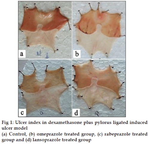
Wistar rats of either sex were divided into four groups of 6 animals each. In this method rats were fasted in individual cages for 24 h. Care was taken to avoid coprophagy. Control vehicle, omeprazole (20 mg/ kg), rabeprazole (20 mg/kg) and lansoprazole (20 mg/ kg) were administered by oral route 30 min prior to ligation. Dexamethasone suspended in 1% w/v CMC was given orally to all the rats 15 min after pylorus ligation. Under ether anesthesia, the abdomen was opened and the pylorus was ligated [14]. The abdomen was then sutured and animals were allowed to recover from the anesthesia. The animals were sacrificed after 4 h, stomachs were dissected out and gastric contents were drained into tubes and subjected to estimation for pH, free and total acidity [15], total proteins [16], pepsin [17] and glandular portions for mucin [18]. The stomachs were then cut open along the greater curvature and the severity of hemorrhagic erosions in the acid secreting mucosa was assessed on a scale of 0 to 3 and ulcer index was determined. Based on their intensity, the ulcers were given scores as follows; 0 indicates no ulcer, 1 denotes superficial mucosal erosion, 2 indicates deep ulcer or transmural necrosis and 3 denotes perforated or penetrated ulcer. The ulcer index was determined using the formula [19], Ulcer index= 10/X, where X is the total mucosal area/total ulcerated area. The results obtained from the present study were analyzed using one-way ANOVA followed by Dunnett’s multiple comparison test using GraphPad Prism 5. The results were expressed as the mean±SEM.
The PPIs significantly reduced the gastric volume, total and free acidity, and increased the pH of the gastric fluid, proving its antisecretory activity. Animals in the omeprazole, rabeprazole and lansoprazole groups showed decrease in volume of gastric juice by 41.17, 48.46 and 34.31 %, respectively, free acidity was found to be 51.13, 63.44 and 30.60 %, respectively and total acidity was found to be 47.65, 55.16 and 35.07 %, respectively. pH was found to increase by 148.87, 166.85 and 134.83 %, respectively. It is evident from the results that rabeprazole is more effective than omeprazole and lansoprazole (Table 1).
| Group | Treatment | Volume of gastric juice (ml) | pH | Free acidity (Meq/100 g) | Total acidity (Meq/100 g) |
|---|---|---|---|---|---|
| I | Control | 11.90± 0.11 | 1. 78±0.09 | 44.6 7±0.49 | 88.83± 0.87 |
| II | Omeprazole | 7.00±0 .27a | 4.43±0.10a | 21.83±0.95a | 46.50±2.26a |
| III | Rabeprazole | 6.13±0.27a | 4.75±0.09a | 16.33±1.26a | 39.83±1.54a |
| IV | Lansoprazole | 7.82±0.22a | 4.18±0.16a | 31.00±1.37a | 57.67±2.47a |
Values are mean±SEM; n=6. ap<0.01 when compared to control group.
Table 1: Effect of proton pump inhibitors on volume of gastric juice, ph, free acidity and total acidity
Percent protection in ulcer index offered by omeprazole, rabeprazole and lansoprazole was 83.92, 89.28, and 79.45, respectively (Table 2). Rabeprazole was found to be most effective. There was significant increase in mucin and reduction in protein and pepsin content in PPIs treated groups compared to control group (Table 3).
| Group | Treatment | Ulcer index | Protection (%) |
|---|---|---|---|
| I | Control | 9.33±0.25 | --- |
| II | Omeprazole | 1.50±0.22a | 83.92 |
| III | Rabeprazole | 1.00±0.20a | 89.28 |
| IV | Lansoprazole | 1.92±0.37a | 79.45 |
Values are mean±SEM; n=6. ap<0.01 when compared to control group.
Table 2: Effect of proton pump inhibitors on ulcer index
| Group | Treatment | Mucin content (μg/g of wet gland) | Total proteins (μg/ml) | Pepsin (μg/ml) |
|---|---|---|---|---|
| I | Control | 216.7±1.453 | 355.3±1.856 | 15.02±0.691 |
| II | Omeprazole | 242.3±3.266a | 309.2±5.974a | 7.5±0.1706a |
| III | Rabeprazole | 255.8±5.608a | 280.0±3.941a | 6.6±0.5258a |
| IV | Lansoprazole | 236.8±4.400b | 321.7±6.540a | 8.8±0.2352a |
Values are mean±SEM; n=6. ap<0.001 when compared to control group. bp<0.01 when compared to control group.
Table 3: Effect of proton pump inhibitors on mucin, total proteins and pepsin
In the present study gastric ulcer was induced by dexamethasone plus pylorus ligation model. Pylorus ligation induced ulcers are due to autodigestion of the gastric mucosa and break down of the gastric mucosal barrier. Reactive oxygen species are involved in the pathogenesis of pylorus ligation induced mucosal injury [20]. Gastric ulcer and gastrointestinal bleeding are recognized complications of corticosteroid therapy. It causes ulcers by increase in the generation of lipid peroxides indicating the involvement of free radicals in the process of ulceration [21]. Ulcerogenic potential of corticosteroids is well known and is believed to be a result of increased gastric acid secretion and decrease in PGs synthesis. It also delays the gastric ulcer healing by inhibiting epithelial cell proliferation and angiogenesis at the ulcer margin. In dexamethasone plus pylorus ligation model, PPIs viz. omeprazole, rabeprazole and lansoprazole significantly decrease the gastric juice volume, acid and pepsin output indicating decrease in offensive acid and pepsin secretion. On the defensive factors, PPIs significantly increased the gastric mucin secretion and prevented the gastric mucosal damage induced by dexamethasone plus pylorus ligation. Among these PPIs, rabeprazole showed better reduction of gastric acid secretion and decrease in ulcer index than to omeprazole and lansoprazole. This effect of rabeprazole may be due to rapid onset of H+/ K+ ATPase pump inhibition and a greater effect on intragastric pH as compared to omeprazole and lansoprazole. The present findings agree with earlier reports.
Omeprazole, rabeprazole and lansoprazole have shown increased gastric pH and decrease in the protein content of gastric juice. Gastric acid is an important factor for the genesis of ulceration of pylorus ligation ulcer in rats. Gastric acid secretion is influenced by many factors including anxietic effect in the CNS, vagal activity, cholinergic, histaminergic and gastrinergic transmissions [22]. The antisecretory actions of PPIs mainly involve inhibition of H+/K+ ATPase pump. Cytoprotective effect of omeprazole is due to increased expression of COX-2 protein and elevating the levels of PGE2. It also showed increased gastric pH and reduction in gastric acid secretion, which may be due to inhibition of gastric mucosa enzymes, carbonic anhydrase II (CA) and CA IV, which are located in abundance in the gastric parietal cells and in the secretory canaliculi walls. This inhibition potentiates the inhibitory effect on the proton pump [23]. Similarly lansoprazole exerts its gastroprotective effects by increased bio-availability of mucosal sulfhydryl compounds and possibly prostaglandins [24].
Amount of gastric mucus secretion in gastric mucosa was found to be increased in rabeprazole treated group [25]. This mucus consists of mucin-type glycoproteins, which can be detected by amount of alcian blue binding [26] (fig. 1). The decrease in the protein content of gastric juice by rabeprazole suggests the decrease of leakage of plasma proteins into gastric juice. This further suggests the increase in glycoprotein content of the gastric mucosa and that acts as a coating as well as protective barrier on the mucosa.
Thus, the ulcer healing effect of rabeprazole may be due to its effect on both offensive and defensive factors. Further work on other mucosal factors like nitric oxide, prostaglandins, cAMP etc would provide more insight into the activity of rabeprazole.
Acknowledgements
The authors thank Cadila healthcare, Ahmedabad and Cipla Laboratories, Goa for providing the drug samples. The authors are grateful to the Principal, K. L. E. S College of Pharmacy, Hubli for providing the necessary facilities to carry out the work.
References
- Bernersen B, Johnsen R, Straume B, Burhol PG, Jennsen TG, Stakkevold PA. Towords a true prevalence of peptic ulcer: the Sorreisa gastrointestinal disorder study. Gut 1990;31:989-92.
- Friedman JD, Haile PM, Siegelaaub AB, Seltzer CE. Cigarette, alcohol, coffee and peptic ulcer. N Engl J Med 1974;290:469-73.
- Manjari V, Das UN. Oxidant stress, anti-oxidants, nitric acid and essential fatty acids in peptic ulcer disease. Prostaglandins, LeukotEssent Fatty Acids 1998;59:401-6.
- Megraud F, Lamouliatte H. Helicobacter pylori and duodenal ulcer evidence suggesting causation. Dig Dis Sci 1992;5:769-72
- Dujovne CA, Azarnoff DL. Clinical complications of corticosteroid therapy. Med Clin North Am 1973;57:1331-42.
- Tripathi KD. Essentials of Medical Pharmacology. 5thed. New Delhi: Jaypee brothers; 2004. p. 255-65.
- Pezner RD, Lipsett JA. Peptic ulcer disease and other complications in patients receiving dexamethasone palliation for brain metastasis. West J Med 1982;137:375-78.
- Mccall TB, Palmer RM, Moncada S. Induction of nos in rat peritoneal neutrophils and its inhibition by dexamethasone. Eur J Immunol 1991;21:2523-27.
- Bandyopadhyay U, Biswas K, Bandyopadhyay D, Ganguly CK, Banerjee RK. Dexamethasone Makes The Gastric Mucosa Susceptible To Ulceration By Inhibiting Prostaglandin Synthetase And Peroxidase - Two Important Gastroprotective Enzymes. Mol Cell Biochem 1999;202:31-6.
- Wandall JH. Effects of omeprazole on neutrophil chemotaxis, superoxide production degranulation and translocation of cytochrome b-245. Gut 1992;33:617-21.
- Dharmani P, Chauhan Singh V, Palit G. Cyclo oxygenase-2 expression and prostaglandin E2 production in experimental chronic gastric ulcer healing. Eur J Pharmacol 2005;519:277-84.
- Natale G, Lazzeri G, Lubrano V, Colucci R, Vassalle C, Fornai M, etal. Mechanisms of gastroprotection by lansoprazole pretreatment againstexperimentally induced injury in rats role of mucosal oxidative damage and sulfhydryl compounds. ToxicolApplPharmacol 2004;195:62-72.
- John H. The proton pump inhibitors: Similarities and differences. ClinTher 2000;22:266-80.
- Shay M, Komarov SA, Fels D, Meranze D, Gruenstein H.A simple method for the uniform production of gastric ulceration in the rat. Gastroenterology 1945;5:43-61.
- Parmar NS, Hsnnings G, Gulati OP. The gastric antisecretory activity of 3-methoxy-5,7,3’,4’tetrahydroxyflavan (ME)-a specific histidine decarboxylase inhibitor in rats. Agents Actions 1984;15:143-5.
- Lowry OH, Rosebrough NJ, Farr AL, Randall RJ. Protein measurement with the folin phenol reagent. J BiolChem 1951;193:265.
- Anson ML. Estimation of pepsin, trypsin, papain and cathepsin with hemoglobin. J Gen Physiol 1938;22:79-89.
- Come SJ, Morrisey SM, Woods KJ. A method for the quantitative estimation of gastric barrier mucus. J Physiol 1974;2452:116-7.
- Ganguly AK. A method for quantitative assement of experimentally produced ulcers in stomach of rats. Experientia 1969;25:1124.
- Rastogi L, Patnaik GK, Dikshit M. Free radicals and antioxidant status following pylorus ligation induced gastric mucosal injury in rats. Pharmacol Res 1998;38:2125-132.
- Manjari V, Das UN. Effect of polyunsaturated fatty acids on dexamethasone induced gastric mucosal damage. Prostaglandins LeukotEssent Fatty Acids 2000;2:85-96.
- Rang HP, Dale MM, Ritter JM, Flower RJ. Rang and Dale’s Pharmacology. 6thed. New Delhi: Elsevier; 2003. p. 367-72.
- Puscas I, Coltau M, Baican M, Domuta G. Omeprazole has dual mechanism of action. It inhibits both H+K+ ATPase and gastric mucosa carbonic anhydrase enzyme in humans (in vitro and in vivo experiments). J PharmacolExpTher 1999;290:530-4.
- Blandizzi C, Natale G, Gherardi G, Lazzeri G, Marveggio MC, Colucci R, Carignani D, et al. Acid-independantgastroproptective effects of lansoprazole in experimental mucosal injury. Dig Dis Sci 1999;24:2039-50.
- Skoczylas T, Sarosiek I, Sostarich S, McElhinney C, Durham S, Sarosiek J. Significant enhancement of gastric mucin content after rabeprazole administration: its potential clinical significance in acid-related disorders. Dig Dis Sci 2003;48:322-8.
- Clamp JR, Roth J, Lucocq JM, Berger EC, Pantson JC, Watkins WM. Chemical aspects of mucus. Br Med Bull 1978;34:25-41.
