- *Corresponding Author:
- Reema Gabrani
Department of Biotechnology, Jaypee Institute of Information Technology, Noida-201 307, India
E-mail: reema.gabrani@jiit.ac.in
| Date of Submission | 24 January 2012 |
| Date of Revision | 31 August 2012 |
| Date of Acceptance | 2 September 2012 |
| Indian J Pharm Sci 2012, 74 (5): 451-453 |
Abstract
Solanum nigrum is used in various traditional medical systems for antiproliferative, antiinflammatory, antiseizure and hepatoprotective activities. We have evaluated organic solvent and aqueous extracts obtained from berries of Solanum nigrum for antiproliferative activity on leukemic cell lines, Jurkat and HL-60 (Human promyelocytic leukemia cells). The cell viability after the treatment with Solanum nigrum extract was measured by MTT (3-[4,5-dimethylthiazol-2-yl]-2,5-diphenyl tetrazolium bromide) assay. Results indicated increased cytotoxicity with increasing extract concentrations. Comparative analysis indicated that 50% inhibitory concentration value of methanol extract is the lowest on both cell lines.
Keywords
Cytotoxicity, HL‑60, Jurkat, MTT assay, Solanum nigrum
Natural products play a very important role in the new drug discovery and development. Many developing countries depend to a large extent on the traditional system of healthcare largely comprising of medicinal plants and their products. But in comparison to the allopathic system, herbal remedies have been scientifically validated to a lesser extent. The knowledge about the bioactivity of phytomedicinal compound, its potential mode of action and target site identification will help in its utilisation as alternative therapeutic agent [1]. Drugs derived from natural products such as morphine, codeine (from opium poppy), digoxin (from digitalis leaves), atropine and hyoscine (from Solanaceae plants) are all clinically used. Solanum nigrum, also belonging to Solanaceae, is widely used in many traditional systems of medicine worldwide for disparate ailments but has recently garnered attention for the modern therapeutic approaches.
S. nigrum is a herbal plant found in most parts of the world. Various epidemiological studies have indicated that S. nigrum protects against various ailments [2]. It has been used traditionally for the treatment of bacterial infections, cough and indigestion. This plant has also been investigated for antiproliferative [3,4], antiseizure [5], antioxidant [6], antiviral [7], antiinflammatory [8] and hepatoprotective effects [9,10].
S. nigrum has been reported to possess various compounds, which are responsible for diverse activities. The main active constituents reported are glycoalkaloids, glycoproteins and polysaccharides [2,3,6]. These phytochemicals are known to possess various activities. Both the crude extract and the isolated components of S. nigrum have been shown to possess antiproliferative activity on various tumour cell lines such as HepG2 [11,12] (liver), HT29 [12] and HCT‑116 [13] (colon), MCF‑7 [14] (breast), U‑14 [15] and HeLa [16] (cervical). It has been reported that biological activity of the extract may vary based on its extraction method [17].
In the present study, we have carried out a preliminary investigation of the antiproliferative effect of the organic and aqueous extracts of S. nigrum on human leukemic cell lines, Jurkat and HL‑60. We found that S. nigrum extract (SNE) reduced cell viability in a dose‑dependent manner.
S. nigrum was collected from a local nursery at Noida, UP, India and was authenticated by comparing the morphological features. It was planted in the Institute’s nursery and was regularly monitored.
Ripe berries collected from S. nigrum plant were crushed into fine powder and 10 g was extracted with methanol and ethanol separately using a Soxhlet apparatus. The collected organic extract was concentrated in a rotary evaporator under reduced pressure.
To prepare aqueous extract, 10 g of the sample powder was suspended in 25 ml of HEPES (4‑(2‑hydroxyethyl)‑1‑piperazineethanesulfonic acid) buffer (pH 8.0) containing 1 mM NaCl as described earlier [18]. The solution was incubated at 25° for 1 h and then filtered using the millipore filter of 0.45 μm. The filtrate was centrifuged following incubation at 90° for 10 min and the supernatant was used.
Jurkat and HL‑60 cell lines were cultured in Roswell Park Memorial Institute (RPMI)‑1640 supplemented with 100 U/ml penicillin, 100 μg/ml streptomycin and 10% fetal bovine serum. All cell cultures were maintained in humidified atmosphere of 5% CO2 at 37°. For cell viability assay, Jurkat and HL‑60 cells were seeded in 96 well tissue culture plates at 1×10⁵ cells/ml and incubated at 37°, 5% CO2 for 24 h to allow the cells to adhere to the plate. Subsequently the cells were treated with different dilutions of the SNE for 48 h at 37° and 10 μl of MTT (5 mg/ml) was added to each well and again incubated for 4 h at 37°. Lastly, the formazan produced was solubilized by adding dimethyl sulphoxide and absorbance was measured at 570 nm in an enzyme‑linked immunoabsorbent assay microplate reader. The readings obtained were used to calculate the cell viability ( [Aexperimental/ Acontrol]×100) and IC50 value [19]. The IC50 value, i.e., the concentration required to inhibit 50% of cells viability was determined by plotting the log of the drug concentration versus the percent inhibition. Best‑fit line was plotted by least‑squares linear regression. The 50% inhibitory concentration (IC50) was calculated from the linear‑regression equation: Log (CV50)=m×log (IC50)+c; where m is regression coefficient, c is intercept of the line, log (IC50) is the log of the 50% inhibitory concentration of the extract and log (CV50) is the log value of 50% cell viability.
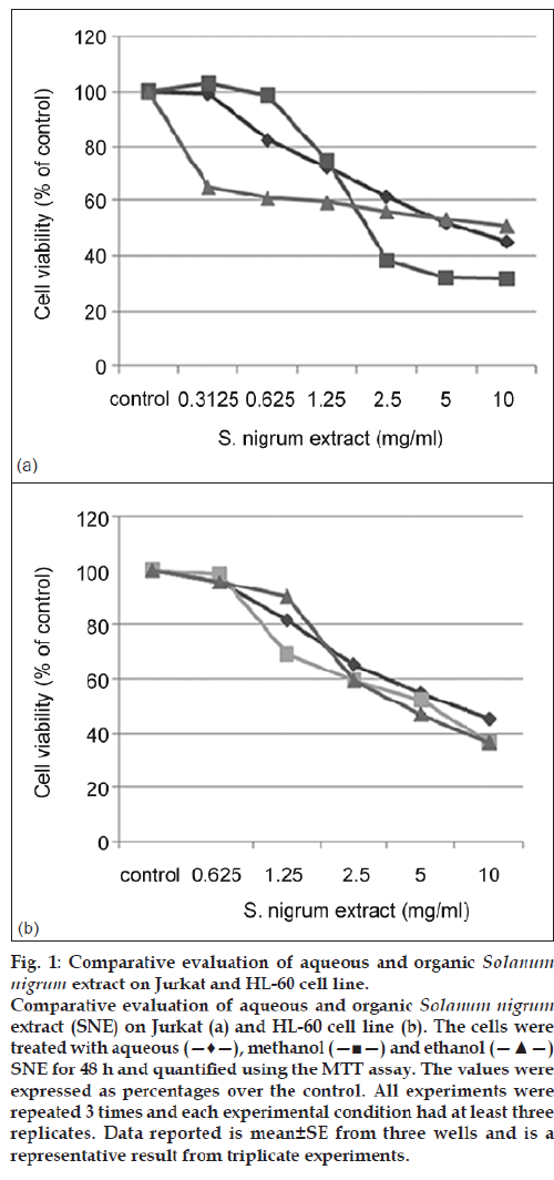
The cytotoxic effects of various concentrations of aqueous and organic extracts of SNE were analysed on leukemic cell lines, Jurkat and HL‑60. Jurkat is acute T‑cell leukemic cell line and HL‑60 is a promyelocytic cell line. Both of these cell lines are of human origin. Jurkat and HL‑60 cells were treated with the increasing concentrations of SNE (methanol, ethanol and aqueous) for 48 h and their viability was evaluated using MTT assay. The cytotoxicity of all three extracts was found to be concentration‑dependent (fig. 1). In another study, the crude extract of S. nigrum was also shown to exert dose‑dependent response in the hepatic cancer cell line HepG2 inducing different responses in the cells at low and high extract concentration. It was reported that the hepatic cells underwent apoptosis upon treatment with higher concentration whereas lower concentration resulted in autophagocytosis [19]. Moreover, another recent study reported the antitumor potential of S. nigrum fruits on HeLa cell line whereas its effect on Vero, normal monkey kidney cell line, was less prominent concluding that this drug has considerable anticervical cancer activity [16].
Figure 1: Comparative evaluation of aqueous and organic Solanum
nigrum extract on Jurkat and HL-60 cell line.
Comparative evaluation of aqueous and organic Solanum
nigrum extract (SNE) on Jurkat (a) and HL-60 cell line (b). The cells were
treated with aqueous (—♦—), methanol (—■—) and ethanol (—▲—)
SNE for 48 h and quantified using the MTT assay. The values were
expressed as percentages over the control. All experiments were
repeated 3 times and each experimental condition had at least three
replicates. Data reported is mean±SE from three wells and is a
representative result from triplicate experiments.
The methanol SNE was more effective as compared to other extracts towards both Jurkat T cell line (IC50: 3 mg/ml) (fig. 1a) and HL‑60 promyelocytic cell line (IC50: 4.8 mg/ml) (fig. 1b); whereas its effect was more pronounced on Jurkat cells. The IC50 value of aqueous SNE was found to be twice that of methanol SNE on Jurkat cells. These results suggest that methanol was more efficient in extracting the active compound from the crude extract as compared to ethanol and water based solvent.
Recently, the antiproliferative effect of S. nigrum has also been shown against various cancer cell lines. The antitumour effect of SNE has been indicated due to the presence of polysaccharide [20], glycoalkaloids [11] or glycoproteins [15,16]. The polysaccharides have been shown to exert antiproliferative effect due to their immunomodulatory properties whereas glycoalkaloids and glycoproteins have been shown to activate the proapoptotic factors or inhibit the transcription factors playing an important role in tumour progression.
Thus the organic and aqueous SNE were cytotoxic in dose dependent manner on two different leukemic cell lines, i.e. Jurkat and HL‑60. The SNE mediated inhibition of cell growth was found to be more pronounced on Jurkat cells as compared to HL‑60 cell line. These studies suggest the beneficial potential of S. nigrum as an antitumour agent although the mechanism of action behind it remains to be elucidated.
References
- Briskin DP. Medicinal plants and phytomedicines. Linking plant biochemistry and physiology to human health. Plant Physiol 2000;124:507-14.
- Jain R, Sharma A, Gupta S, Sarethy IP, Gabrani R. Solanumnigrum : Current perspectives on therapeutic properties. Altern Med Rev 2011;16:78-85.
- Li J, Li QW, Gao DW, Han ZS, Lu WZ. Antitumor and immunomodulating effects of polysaccharides isolated from Solanumnigrum Linne. Phytother Res 2009;23:1524-30.
- Nawab A, Thakur VS, Yunus M, Ali Mahdi A, Gupta S. Selective cell cycle arrest and induction of apoptosis in human prostate cancer cells by a polyphenol-rich extract of Solanumnigrum . Int J Mol Med 2012;29:277-84.
- Wannang NN, Anuka JA, Kwanashie HO, Gyang SS, Auta A. Antiseizure activity of the aqueous leaf extract of Solanumnigrum linn (solanaceae) in experimental animals. Afr Health Sci 2008;8:74-9.
- Lee SJ, Lim KT. Antioxidative effects of glycoprotein isolated from Solanumnigrum Linne on oxygen radicals and its cytotoxic effects onthe MCF-7 cell. J Food Sci 2003;68:466-70.
- Javed T, Ashfaq UA, Riaz S, Rehman S, Riazuddin S. In vitro antiviral activity of Solanumnigrum against hepatitis C virus. Virol J 2011;8:26.
- Kang H, Jeong HD, Choi HY. The chloroform fraction of Solanumnigrum suppresses nitric oxide and tumor necrosis factor-αinLPS-stimulated mouse peritoneal macrophages through inhibition of p38, JNK and ERK1/2. Am J Chin Med 2011;39:1261-73.
- Lin HM, Tseng HC, Wang CJ, Lin JJ, Lo CW, Chou FP. Hepatoprotective effects of Solanumnigrum Linn. extract against CCl4-induced oxidative damage in rats. ChemBiol Interact 2008;171:283-93.
- Hsieh CC, Fang HL, Lina WC. Inhibitory effect of Solanumnigrum on thioacetamide-induced liver fibrosis in mice. J Ethnopharmacol 2008;119:117-21.
- Ji YB, Gao SY, Ji CF, Zou X. Induction of apoptosis in HepG2 cells by solanine and Bcl-2 protein. J Ethnopharmacol 2008;115:194-202.
- Lee KR, Kozukue N, Han JS, Park JH, Chang EY, Baek EJ, et al. Glycoalkaloids and metabolites inhibit the growth of humancolon (HT29) and liver (HepG2) cancer cells. J Agric Food Chem 2004;52:2832-9.
- Lee SJ, Oh PS, Ko JH, Lim K, Lim KT. A 150-kDa glycoprotein isolated from Solanumnigrum L. has cytotoxic and apoptotic effects by inhibiting the effects of protein kinase C alpha, nuclear factor-kappa B and inducible nitric oxide in HCT-116 cells. Cancer ChemotherPharmacol 2004;54:562-72.
- Son YO, Kim J, Lim JC, Chung Y, Chung GH, Lee JC. Ripe fruit of Solanumnigrum L. inhibits cell growth and induces apoptosis in MCF-7 cells. Food Chem Toxicol 2003;41:1421-8.
- Li J, Li Q, Feng T, Li K. Aqueous extract of Solanumnigrum inhibit growth of cervical carcinoma (U14) via modulating immune response of tumor bearing mice and inducing apoptosis of tumor cells. Fitoterapia 2008;79:548-56.
- Oh PS, Lim KT. HeLa cells treated with phytoglycoprotein (150 kDa) were killed by activation of caspase 3 via inhibitory activities of NF-kappaB and AP-1. J Biomed Sci 2007;14:223-32.
- Das K, Tiwari RK, Shrivastava DK. Techniques for evaluation of medicinal plant products as antimicrobial agent: Current methods and future trends. J Med Plants Res 2010;4:104-11.
- Liu Y, Luo J, Xu C, Ren F, Peng C, Wu G, et al. Purification, characterization, and molecular cloning of the gene of a seed-specific antimicrobial protein from pokeweed. Plant Physiol 2000;122:1015-24.
- Lin HM, Tseng HC, Wang CJ, Chyau CC, Liao KK, Peng PL, et al. Induction of autophagy and apoptosis by the extract of Solanumnigrum Linn in HepG2 cells. J Agric Food Chem 2007;55:3620-8.
- Li J, Li Q, Peng Y, Zhao R, Han Z, Gao D. Protective effects of fraction 1a of polysaccharides isolated from Solanumnigrum Linne on thymus in tumor-bearing mice. J Ethnopharmacol 2010;129:350-6.
